Abstract
- The precise mechanism involved in DIC and delayed traumatic subarachnoid hemorrhage (DT-SAH) remains unclear in multipletrauma patients. Hereby, we describe a polytraumatized patient with DIC who died due to DT-SAH. A 75-year-old female patient was admitted to our Emergency Department complaining of abdominal pain and drowsiness after a pedestrian accident. Her initial brain computerized tomography (CT) finding was negative for intracranial injury. However, her abdominal CT scan revealed a collection of retroperitoneal hematomas from internal iliac artery bleeding after a compressive pelvic fracture. This event eventually resulted in shock and DIC. An immediate angiographic embolization of the bleeding artery was performed along with transfusion and antithrombin III. Her vital signs were stabilized without neurological change. Fourteen hours after admission, she suddenly became comatose, and her follow-up brain CT scan revealed a dense DT-SAH along the basal cisterns with acute hydrocephalus. This event rapidly prompted brain CT angiography and digital subtraction angiography, which both confirmed the absence of any cerebrovascular abnormality. Despite emergency extraventricular drainage to reverse the hydrocephalus, the patient died three days after the trauma. This paper presents an unusual case of DT-SAH in a polytraumatized patient with DIC.
-
Keywords: disseminated intravascular coagulation; polytrauma; taumatic subarachnoid hemorrhage
Disseminated intravascular coagulation (DIC) is commonly associated with multiple trauma. Severe head injuries also reported for decades to cause DIC with the release of brain thromboplastin as a high risk factor of systemic blood coagulopathy. [1,2] The concept of delayed traumatic intracerebral hemorrhage (DT-ICH) was first reported as “Traumatische Spät-Apoplexie” by Bollinger in 1891.[3] Many reports along with advances in computerized tomography (CT) techniques redefined the meaning of DT-ICH based on radiologic findings of newly formed hematomas in follow-up studies in which the initial CT scan was normal. However, most literatures described DT-ICH as an intra-parenchymal hemorrhage as a part of hematoma expansion rather than a newly developed hematoma formation due to the ill-defined characterization of DT-ICH. The DT-ICH terminology is also rather mixed, containing a wide range of hemorrhages, such as DT- epidural hematomas, intracranial hemorrhages, and subarachnoid hemorrhage (SAH). Among these, the DT-SAH was considered a secondary phenomenon of hemorrhage leakage from the existing intra-parenchymal hemorrhage or subdural hematoma. To our knowledge, this is a unique case of a polytraumatized patient presenting with DT-SAH when her initial brain CT scan for trauma evaluation was radiologically normal. Here, the authors share the experience of a patient who eventually expired due to the complications of DT-SAH following DIC.
Case Report
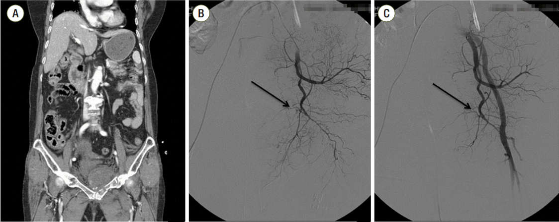
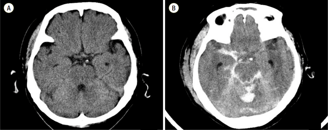
A 75-year-old woman was admitted for abdominal pain after a pedestrian traffic accident. The patient was diabetic but had no other medical history such as anticoagulation therapy. Her initial mental status was drowsy, and her Glasgow Coma Scale (GCS) score was 13. On the other hand, her initial blood pressure 86/53 mmHg while her heart rate was 142 beats per minute. One hour after the accident, the initial CT brain scan was performed. Her initial CT showed no definite hemorrhagic lesion (Fig. 1). She was referred to the Department of Trauma Surgery for a pelvic bone fracture and retroperitoneal hematoma management. A CT of her abdomen and pelvis showed a large retroperitoneal hematoma due to a left iliac artery injury. Her initial hemoglobin level was 7.6 g/dL. In spite of transfusion of 2 packs of red blood cells, her blood pressure was dropped to 69/50 mmHg and the follow-up hemoglobin level was 6.7 g/dL (Table 1). Therefore, emergent pelvic angiography was done. On pelvic angiography, contrast extravasation was revealed at the distal part of the internal iliac artery. Thus, we performed embolization with Spongostan standard®MS0002 (Johnson and Johnson, New Jersey, United States) (Fig. 2). The blood pressure checked before the pelvic embolization was 80/60 mmHg whereas the blood pressure checked three hours after pelvic embolization was recovered up to 129/61 mmHg. Unfortunately, DIC was diagnosed based on her initial lab results taken at the Emergency Department (Table 1). To stabilize the abnormal hematologic factors, the patient was given a transfusion of antithrombin III (500 IU, Green Cross, Yongin, Korea), six 400-mL packs of packed red cell, eight 400-mL packs of platelet concentrate, and eight 400-mL packs of Fresh Frozen plasma (FFP) in one day.
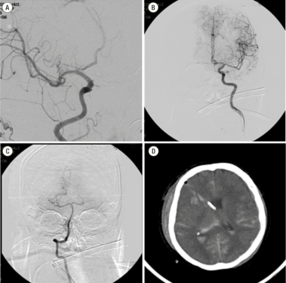
Fourteen hours after the initial CT, the patient’s mental status changed from drowsy to coma. The emergent CT brain was checked, and it showed a newly developed all cisternal SAH and intraventricular hemorrhage (Fig. 1). Because of the aneurismal SAH possibility, we performed digital subtraction angiography and brain CT angiography (CTA). No definite aneurismal lesion was confirmed. For intracranial pressure (ICP) monitoring and cerebrospinal fluid (CSF) drainage, we performed an extraventricular drainage (EVD) procedure (Fig. 3). At the time of ventricular puncture, the CSF was gushed out through the catheter and high intracranial pressure was measured using the calibrated EVD catheter. The measured ICP was over 250 mm CSF which was above the measuring capacity of calibrated EVD catheter. As a high ICP was checked on site, the manometer was not used and the ICP was only measured by the calibrated EVD catheter. The next day, the patient’s pupils were fully dilated, and lung congestion had developed. Three days from admission, the patient expired.
Discussion
This is a report of a mortality case in which a patient died of DT-SAH after multiple trauma. To be more specific, her initial brain CT scan was radiologically normal, but she suffered from DIC shortly after massive abdominal bleeding, and this presumably caused DT-SAH, leading to death in the patient. Before we analyze this case, we must set the definitions of DT-ICH and DT-SAH. DT-ICH refers to a newly formed hematoma found in follow-up studies when the initial brain CT scan is normal. Although this is the theoretical definition, it is somewhat accepted to include a wide range of hemorrhages (i.e., not only newly developed hematomas but also hematoma expansion).[4] Moreover, in the literature, DT-ICH truly means intra-parenchymal hemorrhage in literature, but in the field, it includes most types of intracranial hemorrhage, such as subdural hematomas, epidural hematomas, and SAH. Naturally, DT-SAH is more or less a subset of DT-ICH in this case. Hence, there are many reports on DT-ICH with this broad definition; however, there has been no specific report on pure DT-SAH, such as this report.
The mechanisms of DT-SAH are understood through three classic theories. The first, the theory of necrotic brain softening, was suggested by Bollinger.[3] Necrotic brain softening adjacent to the vessels causes vascular injury, leading to hematoma formation. Secondly, von Holder[5] reported a theory of coalescence of extravasated micro-hematomas. The local cerebral and vascular injury causes blood to leak out from the vessels, which consequently activates hematoma expansion. Thirdly, the theory of dysregulation was introduced to explain the cerebral loss of autoregulation of the brain vessel by hypotension and hypoxia that eventually leads to vessel dilatation and increase of intracranial pressure (IICP).[6] This pathological sequence results in hemorrhagic events. All three theories are commonly accepted, but with DT-SAH, one must consider one more aspect: ruling out the possibility of aneurysmal rupture. A cerebral aneurysm is defined as a cerebrovascular disorder in which weakness in the wall of a cerebral artery causes the localized dilation or ballooning of the blood vessel. As in our case, all cisternal SAHs have the typical radiological features of spontaneous SAHs. Thus, we performed a routine CTA and digital subtraction angiography and found no definite aneurysmal sac. However, we had to consider the possibility that we could not see the aneurysmal sac due to high ICP leading to a false negative result.
Although there are few reports on DT-SAH, when considering the fact that traumatic SAH has been reported to be one of the poor prognostic predictors in traumatic brain injury (TBI) compared to other intracranial hemorrhages, it is generally understood that DT-SAH has a poorer prognosis than parenchymal or extra-parenchymal hemorrhagic lesions.[7,8] Depending on the location and volume of the SAH, the prognosis can vary among DT-SAH patients, but DT-SAH is usually due to arterial bleeding; thus, it is also thought to have a poorer prognosis. Spontaneous SAH has a mortality rate of 40-50%, and it is well known to be poorer than that of Spontaneous intracerebral hemorrhage. This is partly because almost all cisternal SAH is caused by arterial bleeding, thus, arterial rupture may directly damage the brain, resulting in IICP with lower brain perfusion.[9]
There is a definite clinical difference between DT-SAH and other types of DT-ICH. Traumatic intracerebral hemorrhage (T-ICH) and traumatic subarachnoid hemorrhage (TSAH) are categorized as intracranial hemorrhage. However, the evaluation process, treatment plan, and prognosis of the diseases after diagnosis are clinically different due to their distinctive natural course. Obviously, the diagnosis and clinical presentation of other kinds of delayed T-SAH and T-ICH are very similar. These disease entities are clinically presented similarly with symptoms such as sudden mental deterioration and the neurological changes. They are also diagnosed mostly by brain CT scans. However, the evaluation process and the management strategies are completely different after the onset of the disease. To be more specific, most of DT-ICH is immediately accompanied by mental deterioration, which is dependent on the extent of hematoma expansion, and this event is usually followed by an emergency operation which will subsequently determine the recovering state of the patient. In such cases, simple brain CT scans are mainly fast diagnostic tools for evaluation. On the other hand, in the cases of DT-SAH, the mental state and the prognosis of a patient are determined at the point of occurrence of DT-SAH where the surgical evacuation of hematoma is usually not feasible neuro-anatomically. Plainly speaking, the prevention of its recurrence is the only way of treatment as there is no surgical option. Additionally, the brain CT angiography with contrast and digital subtraction angiography are usually required in the patients with DT-SAH to study the vascular anatomy of the injured brain. For this reason, there is a clear clinical difference between DT-SAH and other intracranial hemorrhages.
Delayed traumatic hemorrhage such as DT-SAH has a typical clinical feature, in that the neurologic deficit and mental changes come after a certain period of time. DT-SAH including DT-ICH comprises 0.3-1.7% of all TBIs with a low incidence rate.[10,11] It is not currently feasible to predict potential hemorrhagic lesions in advance clinically; therefore, the ultimate management is by close monitoring of patients.[12] Early and prompt management is directly linked with better prognostic outcomes for patients. As mentioned before, the intracranial mass such as ICH or epidural hematoma can be noticed earlier for surgical intervention, but SAH will have no benefit from the surgery. Surgery only prevents the secondary rupture but usually would not prevent from the consequences of neurological deficits. For DT-SAH, usually a conservative management is currently the only treatment of option. Thus, unlike other DT-ICH, DT-SAH requires early prevention, early intervention, and the correction of risk factors such as DIC.
There are risk factors of DT-SAH such as shorter time interval from the initial CT scan (< 3 hours), multiple trauma, anticoagulant therapy history, low GCS scores, DIC and so on.[12,13] The patient presented in this case had a shorter time interval from the onset of trauma to the initial CT scan and had a risk of DIC. However, the short time interval was an unmodifiable factor. Therefore, the correction of DIC was critical for the survival of the patient.
DIC is commonly accompanied by multiple trauma in patients with brain injury. The risk factors of DIC include blood transfusion reaction, cancer history, pancreatitis, infection in the blood (especially by bacteria or fungus), liver disease history, pregnancy complications, and recent surgery or anesthetic history. As in this present case, a patient with severe tissue injury is also at a high risk of DIC. The most common symptom of DIC is bleeding, which can present in the entire body and not just the area affected by the injury. It comes along with physical signs, such as bruising and decreases in systemic blood pressure. There are many studies on the treatment of DIC, but the most important treatment is the prompt correction of the underlying cause.[14,15] There are other reports about the benefits of the transfusion of recombinant activated factor VII, FFP, or platelet concentration.[16,17] Our specific case initially underwent a process of embolization of the bleeder. The patient was given a transfusion of antithrombin III (500 IU, Green Cross, Yongin, Korea), eight 400-mL packs of platelet concentrate, and eight 400-mL packs of FFP. Nevertheless, the one-day follow-up serum lab results did not show much improvement (Table 1). The patient was not given recombinant factor VII, Protein C concentrate, or heparin. DIC is characterized by a systemic state of consumptive coagulopathy, hence, it will inevitably influence a bleeding risk in other organs in the body. A numerous evidences have reported that there is a higher chance of delayed hemorrhagic lesions in the brain when trauma patients are accompanied by coagulopathy. [18] However, as mentioned previously, two facts must be reminded that: (1) DIC is a hematologically consumptive condition of hemostatic factors; and (2) a delayed traumatic hemorrhages must be preceded by an individual brain injury. Nonetheless, it is acceptable that not all TBI patients with DIC would develop a delayed intracranial hemorrhagic lesion. Based on this rationale, when DIC and DT-SAH are present together, it is rather difficult to determine a direct causal relationship between DIC and DT-SAH in our case of this specific patient. It is justifiable to argue that DIC is one of aggravating factors which play roles in developing delayed cerebral hemorrhages as DIC itself is a systemic condition with a lack of hemostatic factors in the blood. Finally, we think that DT-SAH is not directly caused by DIC, but, DIC is implicated in aggravating DT-SAH in this specific case.
This present case report is rare and is on the patient with initially normal CT brain scan later expired with pure DT-SAH complications. She was undergoing DIC with severe multiple trauma and a high bleeding risk. Traumatic cerebral hemorrhage also has a higher bleeding risk, and when the initial CT brain scan is normal, the sudden neurological aggravation is more unexpected. In addition, if the initial CT brain scan shows abnormal findings, then the follow-up is performed more carefully, but this was not the situation in this case. Traumatic SAH is already a risk factor for mortality and morbidity in patients with multiple trauma. In most cases, the neurological deterioration is irreversible in recovery. In conclusion, in all severe multiple trauma patients, attention must be paid to any unexpected DT-SAH or delayed cerebral hemorrhages. Despite the prompt management of DIC caused by the pelvic bone fracture in this particular case, we report a case of an expired patient with DT-SAH.
NOTES
-
No potential conflict of interest relevant to this article was reported.
Fig. 1.(A) The initial brain computed tomography (CT) images revealed no definite intracranial hemorrhagic lesion. (B) The follow-up brain CT revealed a dense cisternal subarachnoid hemorrhage.

Fig. 2.(A) The initial abdominal computed tomography revealed a pelvic bone fracture with retroperitoneal hematoma. (B) The preembolization pelvic angiography showed an extravasation of the internal iliac artery branch. (C) After embolization, no definite extravasation was confirmed through pelvic angiography.

Fig. 3.(A), (B), (C) According to digital subtraction angiography, no vascular abnormality was present. (D) We performed extraventricular drainage at the right Kocher’s point. The tapping pressure was higher than 250 mm CSF. CSF: cerebrospinal fluid.

Table 1.Laboratory findings of various hematologic factors associated with coagulopathy
|
Hematologic factors |
Initial |
2 hours after admission (after transfusion of 2 PRC) |
6 hours after admission (after transfusion of 6 PRC) |
Normal |
|
Hemoglobin (g/dL) |
7.6 |
6.7 |
9.0 |
13.5-17.0 |
|
Platelet count (E9/L) |
69 |
89 |
77 |
165-360 |
|
Prothrombin time (sec) |
13.6 |
19.6 |
- |
9.5-12.8 |
|
Partial prothrombin time (sec) |
35.0 |
34.4 |
- |
27.9-37.8 |
|
D-dimer (ng/mL) |
57,732 |
11,245 |
- |
< 280 |
|
Antithrombin (% activity) |
56 |
48 |
- |
77-123 |
|
Fibrinogen degradation product (ug/mL) |
47.9 |
52.9 |
- |
< 5 |
|
Fibrinogen (mg/dL) |
206 |
500 |
- |
200-400 |
References
- 1. Pondaag W. Disseminated intravascular coagulation related to outcome in head injury. Acta Neurochir Suppl (Wien) 1979;28:98-102.ArticlePubMed
- 2. Vecht CJ, Sibinga CT, Minderhoud JM. Disseminated intravascular coagulation and head injury. J Neurol Neurosurg Psychiatry 1975;38:567-71.ArticlePubMedPMC
- 3. Bollinger O. Über traumatische Spät-Apoplexie: Ein Beitrag zur Lehre von der Hirnerschütterung. In: Internationale Beiträge zur wissenschaftlichen Medizin; festschrift, Rudolf Virchow gewidmet zur vollendung seines 70. Lebensjahres. In: Virchow R. Berlin, August Hirschwald. 1891, pp 457-70.
- 4. Tsubokawa T. Two types of delayed post-traumatic intracerebral hematoma. Neurol Med Chir (Tokyo) 1981;21:669-75.ArticlePubMed
- 5. von Holder H. Pathologische Anatomie der Gehirnerschutterung Beim Meim Menschen. Stuttgart. J Weise 1904;12:10-6.
- 6. Evans JP, Scheinker IM. Histologic studies of the brain following head trauma; post-traumatic petechial and massive intracerebral hemorrhage. J Neurosurg 1946;3:101-13.ArticlePubMed
- 7. Paiva WS, de Andrade AF, de Amorim RL, Muniz RK, Paganelli PM, Bernardo LS, et al. The prognosis of the traumatic subarachnoid hemorrhage: a prospective report of 121 patients. Int Surg 2010;95:172-6.PubMed
- 8. Chen G, Zou Y, Yang D. The influence of traumatic subarachnoid hemorrhage on prognosis of head injury. Chin J Traumatol 2002;5:169-71.PubMed
- 9. Ishikawa T, Moroi J, Hikichi K, Yoshioka S, Suzuki A. Prognosis of SAH after surgery-Japan and Asia pacific region. Cerebrovascular Diseases 2012;34:12.
- 10. Kaplan M, Ozveren MF, Topsakal C, Erol FS, Akdemir I. Asymptomatic interval in delayed traumatic intracerebral hemorrhage: report of two cases. Clin Neurol Neurosurg 2003;105:153-5.ArticlePubMed
- 11. Sawauchi S, Yuhki K, Abe T. The relationship between delayed traumatic intracerebral hematoma and coagulopathy in patients diagnosed with a traumatic subarachnoid hemorrhage. No Shinkei Geka 2001;29:131-7.PubMed
- 12. Rim BC, Kim ED, Min KS, Lee MS, Kim DH. A clinical analysis of delayed traumatic intracerebral hemorrhage. J Korean Neurosurg Soc 1998;27:1490-9.
- 13. Lipper MH, Kishore PR, Girevendulis AK, Miller JD, Becker DP. Delayed intracranial hematoma in patients with severe head injury. Radiology 1979;133(3 Pt 1):645-9.ArticlePubMed
- 14. Churliaev IuA, Lychev VG, Epifantseva NN, Redkokasha Llu. Treatment of DIC syndrome in patients with severe craniocerebral trauma. Anesteziol Reanimatol 2001;27-9.
- 15. Gando S, Wada H, Thachil J, Scientific and Standardization Committee on DIC of the International Society on Thrombosis and Haemostasis (ISTH). Differentiating disseminated intravascular coagulation (DIC) with the fibrinolytic phenotype from coagulopathy of trauma and acute coagulopathy of trauma-shock (COT/ACOTS). J Thromb Haemost 2013;11:826-35.ArticlePubMed
- 16. Schmid S, Friesenecker B, Lorenz I, Innerhofer P, Koscielny J, Velik-Salchner C, et al. Administration of recombinant activated factor VII (NovoSeven) in three cases of uncontrolled bleeding caused by disseminated intravascular coagulopathy. Clin Appl Thromb Hemost 2007;13:313-7.ArticlePubMed
- 17. Baharvahdat H, Lari MS, Etemadresaee H, Shariati Z. Effect of fresh frozen plasma in prevention of delayed intracerebral hematoma in severe traumatic brain injury. J Neurotrauma 2004;21:1298.
- 18. Stein SC, Young GS, Talucci RC, Greenbaum BH, Ross SE. Delayed brain injury after head trauma: significance of coagulopathy. Neurosurgery 1992;30:160-5.ArticlePubMed
Citations
Citations to this article as recorded by

- Is initial optic nerve sheath diameter prognostic of specific head injury in emergency departments?
Bedriye Müge Sönmez, Emirhan Temel, Murat Doğan İşcanlı, Fevzi Yılmaz, Uğur Gülöksüz, Selçuk Parlak, Özhan Merzuk Uçkun
Journal of the National Medical Association.2019; 111(2): 210. CrossRef
 , Wonyeon Lee, M.D., Ph.D.2, Ji Young Jang, M.D.3
 , Pilyoung Jung, M.D.3, Sohyun Kim, M.D.1, Jongyeon Kim, M.D.1
 , Jinsu Pyen, M.D., Ph.D.1
 , Kum Whang, M.D. Ph.D.1
 , Sungmin Cho, M.D., Ph.D.1


 KSCCM
KSCCM
 PubReader
 ePub Link
 Cite
Cite


