
Articles
- Page Path
- HOME > Acute Crit Care > Volume 30(1); 2015 > Article
-
Original Article
Thoracic Surgery Complications of Central Venous Totally Implantable Access Port: Internal Jugular Versus Subclavian Access - Pil Young Jung, M.D., Hoon Ryu, M.D., Ph.D., Jae Hung Jung, M.D.*, Eunbi Lee, M.D.†, Joong Hwan Oh, M.D., Ph.D.*, Chun Sung Byun, M.D.*, Il Hwan Park, M.D.*
-
Korean Journal of Critical Care Medicine 2015;30(1):13-17.
DOI: https://doi.org/10.4266/kjccm.2015.30.1.13
Published online: February 28, 2015
Department of Surgery, Wonju College of Medicine, Wonju Severance Christian Hospital, Wonju, Korea
*Department of Thoracic and Cardiovascular Surgery, Wonju College of Medicine, Wonju Severance Christian Hospital, Wonju, Korea
��Department of Anesthesiology, Wonju College of Medicine, Wonju Severance Christian Hospital, Wonju, Korea
- Correspondence to: Il Hwan Park, Department of Thoracic and Cardiovascular Surgery, Wonju College of Medicine, Wonju Severance Christian Hospital, 20, Ilsan-ro, Wonju 220-701, Korea Tel: +82-33-741-1341, Fax: +82-33-742-0666 E-mail: nicecs@yonsei.ac.kr
• Received: November 5, 2014 • Revised: December 18, 2014 • Accepted: December 29, 2014
Copyright © 2015 The Korean Society of Critical Care Medicine
This is an Open Access article distributed under the terms of the Creative Commons Attribution Non-Commercial License (http://creativecommons.org/licenses/by-nc/4.0/) which permits unrestricted non-commercial use, distribution, and reproduction in any medium, provided the original work is properly cited.
- 7,423 Views
- 93 Download
- 1 Crossref
This article has been corrected. See "Complications of Central Venous Totally Implantable Access Port: Internal Jugular Versus Subclavian Access" in Volume 30 on page 365.
Abstract
-
Background
- Totally implantable access port (TIAP) provides reliable, long term vascular access with minimal risk of infection and allows patients normal physical activity. With wide use of ports, new complications have been encountered. We analyzed TIAP related complications and evaluated the outcomes of two different percutaneous routes of access to superior vena cava.
-
Methods
- All 172 patients who underwent port insertion with internal jugular approach (Group 1, n = 92) and subclavian approach (Group 2, n = 79) between August 2011 and May 2013 in a single center were analyzed, retrospectively. Medical records were analyzed to compare the outcomes and the occurrence of port related complications between two different percutaneous routes of access to superior vena cava.
-
Results
- Median follow-up for TIAP was 278 days (range, 1-1868). Twenty four complications were occurred (14.0%), including pneumothorax (n = 1, 0.6%), migration/malposition (n = 4, 2.3%), pinch-off syndrome (n = 4, 2.3%), malfunction (n = 2, 1.1%), infection (n = 8, 4.7%), and venous thrombosis (n = 5, 2.9%). The overall incidence was 8.7% and 20.3% in each group (p = 0.030). Mechanical complications except infectious and thrombotic complications were more often occurred in group 2 (p = 0.033). The mechanical complication free probability is significantly higher in group 1 (p = 0.040).
-
Conclusions
- We suggest that the jugular access should be chosen in patients who need long term catheterization because of high incidence of mechanical complication, such as pinch-off syndrome.
Introduction
Materials and Methods
Results
Discussion
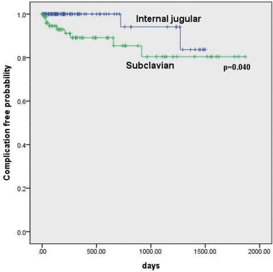
Table 1.Baseline characteristics of patients
| Group 1 | Group 2 | Total | |
|---|---|---|---|
| Total patients, No. (%) | 92 (53.8) | 79 (46.2) | 171 (100) |
| Age (year) | |||
| Median | 62 | 57 | 59 |
| Range | 1-82 | 15-77 | 1-82 |
| Gender, No. (%) | |||
| Male | 34 (37.0) | 24 (30.4) | 58 (33.9) |
| Female | 58 (63.0) | 55 (69.6) | 113 (66.1) |
| Type of disease, No. (%) | |||
| Malignancy | 85 (92.4) | 77 (97.5) | 162 (94.7) |
| Brain | 0 (0) | 1 (1.3) | 1 (0.6) |
| Head and neck | 1 (1.1) | 1 (1.3) | 2 (1.2) |
| Breast | 18 (19.6) | 22 (27.8) | 40 (24.7) |
| Lung | 16 (17.4) | 4 (5.0) | 20 (12.3) |
| Thymus | 0 (0) | 1 (1.3) | 1 (0.6) |
| Esophagus | 1 (1.1) | 4 (5.0) | 5 (3.0) |
| Stomach | 13 (14.1) | 17 (21.5) | 30 (18.5) |
| Colon | 0 (0) | 2 (2.5) | 2 (1.2) |
| Genital | 6 (6.5) | 5 (6.3) | 11 (6.8) |
| Bone | 3 (3.3) | 3 (3.8) | 6 (3.7) |
| Leukemia | 0 (0) | 2 (2.5) | 2 (1.2) |
| Lymphoma | 27 (29.3) | 15 (19.0) | 42 (25.9) |
| Benign | 7 (7.6) | 2 (2.5) | 9 (5.3) |
| Side, No. (%)* | |||
| Right | 81 (88.0) | 20 (25.3) | 101 (59.1) |
| Left | 11 (12.0) | 59 (74.7) | 70 (40.9) |
Table 2.Comparison of port related complications
- 1. Ruesch S, Walder B, Tramèr MR. Complications of central venous catheters: internal jugular versus subclavian access-a systematic review. Crit Care Med 2002;30:454-60.ArticlePubMed
- 2. Galloway M. Insertion and placement of central catheters in the oncology patient. Semin Oncol Nurs 2010;26:102-12.ArticlePubMed
- 3. Broviac JW, Cole JJ, Scribner BH. A silicone rubber atrial catheter for prolonged parenteral alimentation. Surg Gynecol Obstet 1973;136:602-6.PubMed
- 4. Seiler CM, Frohlich BE, Dorsam UJ, Kienle P, Buchler MW, Knaebel HP. Surgical technique for totally implantable access ports (TIAP) needs improvement: a multivariate analysis of 400 patients. J Surg Oncol 2006;93:24-9.ArticlePubMed
- 5. Frykholm P, Pikwer A, Hammarskjöld F, Larsson AT, Lindgren S, Lindwall R, et al. Clinical guidelines on central venous catheterisation. Swedish Society of Anaesthesiology and Intensive Care Medicine. Acta Anaesthesiol Scand 2014;58:508-24.ArticlePubMed
- 6. Silberzweig JE, Sacks D, Khorsandi AS, Bakal CW, Society of Interventional Radiology Technology Assessment Committee. Reporting standards for central venous access. J Vasc Interv Radiol 2003;14(9 Pt 2):S443-52.ArticlePubMed
- 7. Biffi R, Orsi F, Pozzi S, Pace U, Bonomo G, Monfardini L, et al. Best choice of central venous insertion site for the prevention of catheter-related complications in adult patients who need cancer therapy: a randomized trial. Ann Oncol 2009;20:935-40.ArticlePubMed
- 8. Wu CF, Ko PJ, Wu CY, Liu YH, Kao TC, Yu SY, et al. A single-center study of vascular access sites for intravenous ports. Surg Today 2014;44:723-31.ArticlePubMed
- 9. Shin BS, Ahn M. Implantable central venous chemoport: comparision of results according to approach routes and methods. J Korean Radiol Soc 2003;49:165-71.Article
- 10. Araújo C, Silva JP, Antunes P, Fernandes JM, Dias C, Pereira H, et al. A comparative study between two central veins for the introduction of totally implantable venous access devices in 1201 cancer patients. Eur J Surg Oncol 2008;34:222-6.ArticlePubMed
- 11. Silas AM, Perrich KD, Hoffer EK, McNulty NJ. Complication rates and outcomes of 536 implanted subcutaneous chest ports: do rates differ based on the primary operator’s level of training? Acad Radiol 2010;17:464-7.ArticlePubMed
- 12. Keum DY, Kim JB, Chae MC. Safety of a totally implantable central venous port system with percutaneous subclavian vein access. Korean J Thorac Cardiovasc Surg 2013;46:202-7.ArticlePubMedPMC
- 13. Schwarz RE, Groeger JS, Coit DG. Subcutaneously implanted central venous access devices in cancer patients: a prospective analysis. Cancer 1997;79:1635-40.ArticlePubMed
- 14. Ge X, Cavallazzi R, Li C, Pan SM, Wang YW, Wang FL. Central venous access sites for the prevention of venous thrombosis, stenosis and infection. Cochrane Database Syst Rev 2012;3:CD004084. ArticlePubMedPMC
- 15. Mirza B, Vanek VW, Kupensky DT. Pinch-off syndrome: case report and collective review of the literature. Am Surg 2004;70:635-44.ArticlePubMedPDF
- 16. Aitken DR, Minton JP. The “pinch-off sign”: a warning of impending problems with permanent subclavian catheters. Am J Surg 1984;148:633-6.ArticlePubMed
- 17. McGee DC, Gould MK. Preventing complications of central venous catheterization. N Engl J Med 2003;348:1123-33.ArticlePubMed
References
Figure & Data
References
Citations
Citations to this article as recorded by 
- Internal jugular vein versus subclavian vein as the percutaneous insertion site for totally implantable venous access devices: a meta-analysis of comparative studies
Shaoyong Wu, Jingxiu Huang, Zongming Jiang, Zhimei Huang, Handong Ouyang, Li Deng, Wenqian Lin, Jin Guo, Weian Zeng
BMC Cancer.2016;[Epub] CrossRef
 PubReader
 ePub Link
 Cite
Cite

