Prognostic significance of respiratory quotient in patients undergoing extracorporeal cardiopulmonary resuscitation in Korea
Article information

Abstract
Background
Respiratory quotient (RQ) may be used as a tissue hypoxia marker in various clinical settings but its prognostic significance in patients undergoing extracorporeal cardiopulmonary resuscitation (ECPR) is not known.
Methods
Medical records of adult patients admitted to the intensive care units after ECPR in whom RQ could be calculated from May 2004 to April 2020 were retrospectively reviewed. Patients were divided into good neurologic outcome and poor neurologic outcome groups. Prognostic significance of RQ was compared to other clinical characteristics and markers of tissue hypoxia.
Results
During the study period, 155 patients were eligible for analysis. Of them, 90 (58.1%) had a poor neurologic outcome. The group with poor neurologic outcome had a higher incidence of out-of-hospital cardiac arrest (25.6% vs. 9.2%, P=0.010) and longer cardiopulmonary resuscitation to pump-on time (33.0 vs. 25.2 minutes, P=0.001) than the group with good neurologic outcome. For tissue hypoxia markers, the group with poor neurologic outcome had higher RQ (2.2 vs. 1.7, P=0.021) and lactate levels (8.2 vs. 5.4 mmol/L, P=0.004) than the group with good neurologic outcome. On multivariable analysis, age, cardiopulmonary resuscitation to pump-on time, and lactate levels above 7.1 mmol/L were significant predictors for a poor neurologic outcome but not RQ.
Conclusions
In patients who received ECPR, RQ was not independently associated with poor neurologic outcome.
INTRODUCTION
The use of extracorporeal cardiopulmonary resuscitation (ECPR), defined as extracorporeal membrane oxygenation (ECMO) implantation during cardiopulmonary resuscitation (CPR) is increasing. Recent data from the Extracorporeal Life Support Organization Registry have demonstrated a more than 20-fold increase in annual ECPR cases from less than 100 in 2009 to more than 2000 in 2021 [1] and the American Heart Association guideline now gives the recommendation to consider ECPR as an alternative method of CPR for patients with reversible causes of cardiac arrest after more than 10 minutes of conventional CPR [2]. With the increasing use of ECPR, it has become crucial to understand proper management to achieve hemodynamic optimization after ECPR to improve outcomes.
One of the most important goals of CPR including ECPR is to preserve higher cortical function of the brain and many studies have been conducted to investigate if markers of tissue hypoxia are correlated with neurologic outcomes in this patient population [3-5]. An oxygen-derived tissue hypoxia marker: lactate has been associated with neurologic outcomes in these patients. [6]. However, lactate levels can increase with stress response, β-2 adrenergic stimulation, or hepatic dysfunction which suggest that elevated lactate level may not always represent deficiency in tissue oxygen delivery [7]. Thus, CO2-derived markers such as the venous-arterial difference in CO2 tension (ΔpCO2) and respiratory quotient (RQ) may be more useful. In hypoxic states, accumulation of proton in tissues may lead to higher CO2 production due to buffering action of carbonic anhydrase to increase ΔpCO2. But, there are studies that indicate ΔpCO2 better represents status of perfusion rather than tissue hypoxia per se [8,9]. In macro- and micro-circulatory failure, RQ can increase through variety of mechanisms including cells converting to anaerobic metabolism to produce the needed energy. The prognostic role of RQ as a CO2-derived tissue hypoxia marker is relatively well-studied in the sepsis [10,11] and conventional CPR literature [12,13], but the role of RQ in patients with ECPR has not been extensively studied. Thus, this study was performed to investigate whether RQ measured immediately following initiation of ECPR can predict adverse neurologic outcomes in these patients.
MATERIALS AND METHODS
Study Cohort
All adult patients (age >18 years) who received ECPR and survived for >24 hours between May 1, 2004, and April 30, 2020, at a tertiary referral hospital in Seoul, Korea were included in the study. Patients who transferred from other hospitals, who failed ECMO implantation and who had insufficient data for calculating RQ were excluded.
This study was performed according to the Helsinki Declaration and approved by the Institutional Review Board of Samsung Medical Center (No. 2020-10-084-001). The Institutional Review Board waived the requirment for informed consent because of the observational nature of the research.
Operation of ECPR
EPCR was defined as both successful venoarterial ECMO implantation and pump-on with cardiac massage during the index procedure in patients with cardiac arrest. When a return of spontaneous circulation (ROSC) was achieved during ECMO cannulation, ECMO implantation process was not interrupted [14]. Detailed information on the procedure and management of ECPR in our institute has been previously reported [3,15]. In brief, ECPR was considered during cardiac arrest, if the cause of the arrest was presumed to be reversible and the ROSC did not occur despite conventional high-quality CPR performed for more than 10 minutes. ECPR was not performed in patients whose life expectancy was limited to less than 6 months, patients with end-stage malignancies, patients with unwitnessed arrest, patients in whom physical activity was limited, patients in whom the airway was not protected during CPR, or patients in whom CPR was prolonged at the time of initial contact for ECMO implantation (>60 minutes) [3,16].
The intensivists determined the target temperature for targeted temperature management (TTM) according to the hospital's protocol. The TTM was operated using surface cooling devices. We used either cooling pads or a commercial temperature regulation system consisting of hydrogel pads (Arctic Sun, Medivance Corp.).
Definitions and Outcomes
Clinical features, including underlying comorbidities, intensive care unit treatment, and ECPR details, were collected retrospectively by medical record review. All data were collected by a trained study coordinator using a standardized case report form. If the same biological markers were measured several times before ECPR initiation, the value measured at the closest time to ECPR initiation was used.
Arterial and venous blood gas samples collected within a 30-minute interval during the first 24 hours after ECPR initiation were used to calculate RQ. The plasma hemoglobin and lactate levels taken closest to the time of venous blood gas sampling were used to calculate and analyze the RQ. The RQ was calculated as the ratio between the ΔpCO2 and the arterial-venous difference in O2 content (Ca-vO2). Specifically, the calculations were performed as follows: ΔpCO2=venous CO2 tension (PvCO2)–arterial CO2 tension (PaCO2); Ca-vO2=arterial O2 content (CaO2)–venous O2 content (CvO2); CaO2=1.34×arterial O2 saturation (SaO2)×plasma hemoglobin+0.003×arterial O2 tension (PaO2); CvO2=1.34×venous O2 saturation (SvO2)×plasma hemoglobin+0.003×venous O2 tension (PvO2) [17]. The time from initiation of chest compressions to the activation time of ECMO pump was defined as CPR to ECMO pump-on time [3,14].
The primary outcome was neurologic status at hospital discharge. Neurologic status was evaluated by the Glasgow-Pittsburgh cerebral performance categories (CPC) scale [18,19]. Good neurologic outcomes were defined as CPC scores of 1 and 2, and poor neurologic outcomes were defined as CPC score of 3, 4, and 5. Patients were graded on the CPC scale by two independent intensivists who reviewed their medical records thoroughly (YIL and JAR). The secondary outcome was the length of hospital stay after ECPR and hospital mortality.
Statistical Analysis
Continuous variables are described as mean±standard deviation or medians with interquartile ranges (IQRs) and categorical variables are presented as numbers and percentages. To compare characteristics and clinical outcomes between two groups, we used the chi-square test or Fisher’s exact test for categorical variables, and Student t-test or Wilcoxon rank-sum test for continuous variables. A logistic regression model was used to estimate the odds ratios for poor neurologic outcomes. Clinical variables obtained at baseline ECPR with a P<0.1 in univariate analyses were included in the multiple logistic regression model for poor neurologic outcomes. Odds ratios with 95% confidence intervals (CIs) were also calculated. The discriminatory power of each hypoxia parameter was assessed by the area under each receiver operating characteristic curve (AUROC). Statistical analysis were performed with IBM SPSS version 25.0 for Windows (IBM Corp.) and MedCalc 19.5.2 (MedCalc Software). For all analyses, a two-tailed test with a P<0.05 was considered statistically significant.
RESULTS
During the study period, 433 patients underwent ECPR and survived over 24 hours. Patients transferred from other hospitals (n=12), unsuccessful ECMO implantation (n=32), and insufficient data for initial RQ calculation (n=234) were excluded. Finally, data from 155 patients who received ECPR were retrieved for analysis (Figure 1).
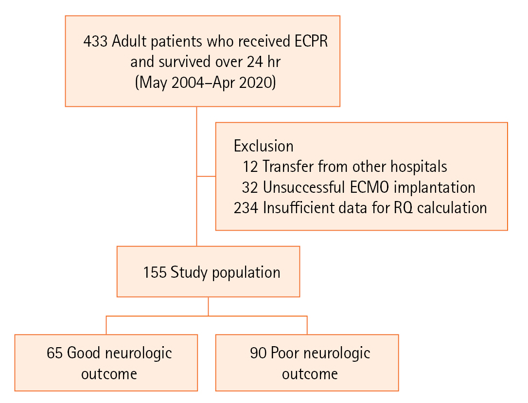
Study flow diagram. ECPR: extracorporeal cardiopulmonary resuscitation; ECMO: extracorporeal membrane oxygenation; RQ: respiratory quotient.
Baseline Characteristics
Baseline clinical characteristics are presented in Table 1. Ninety patients (58.1%) had poor neurologic outcomes at hospital discharge. The median age of the patients was 60.0 (IQR, 51.0–72.0) years, and 112 (72.3%) were males. Sex, body mass index, site of ECMO implantation, initial shockable rhythm, and the proportion of ROSC were similar in both the groups. The proportion of patients with hypertension (54.4% vs. 33.8%, P=0.011) and coronary artery disease (30.0% vs. 15.4%, P=0.035) was higher in the group with the poor neurologic outcome than in the group with good neurologic outcome. Compared with the good neurologic outcome group, the poor neurologic outcome group had a higher proportion of out-of-hospital cardiac arrests (25.6% vs. 9.2%, P=0.010) and longer CPR to pump-on time (33.0 vs. 25.2 minutes, P=0.001).

Baseline characteristics
ECPR Treatment
Data on ECPR management are presented in Table 2. After ECPR, ECMO duration, rate of TTM, rate of mechanical ventilator, vasopressors, and rate of intraaortic balloon counterpulsation did not differ between the two groups. However, the rate of continuous renal replacement therapy (52.2% vs. 23.1%, P<0.001) was higher in the group with poor neurologic outcomes than in the group with good neurologic outcomes. Most ECMO-related complications, such as stroke, limb ischemia, and bleeding at the ECMO site, did not differ between the two groups, but gastrointestinal bleeding (10.0% vs. 1.5%, P=0.046) occurred more frequently in the group with a poor neurologic outcome than in the group with good neurologic outcome.

ECPR treatment
Initial Tissue Hypoxia Parameters and Clinical Outcomes
The number of patients in the two groups according to the distribution of the initial RQ are presented in Figure 2. The median initial RQ of all patients was 2.0 (IQR, 1.4–3.0) (Table 3). The median initial RQ was 2.2 (IQR, 1.5–3.6) in the poor outcome group and 1.7 (IQR, 1.1–2.8) in the good outcome group (P=0.021). In all patients, the median ΔpCO2 was 8.5 mm Hg (IQR, 5.0–14.1 mm Hg), and the median serum lactate level was 7.0 mmol/L (IQR, 3.6–11.7 mmol/L). The median serum lactate level (8.2 vs. 5.4 mmol/L, P=0.004) was elevated in the poor neurologic outcome group than in the good neurologic outcome group; however, the ΔpCO2 value (9.7 vs. 7.0 mm Hg, P=0.093) was similar between the two groups. Patients with poor neurologic outcomes had significantly higher in-hospital mortality (87.8% vs. 0.0%, P<0.001), shorter length of hospital stay after ECPR (5 vs. 22 days, P<0.001), and higher CPC score (5 vs. 1, P<0.001) compared with patients with good neurologic outcome.
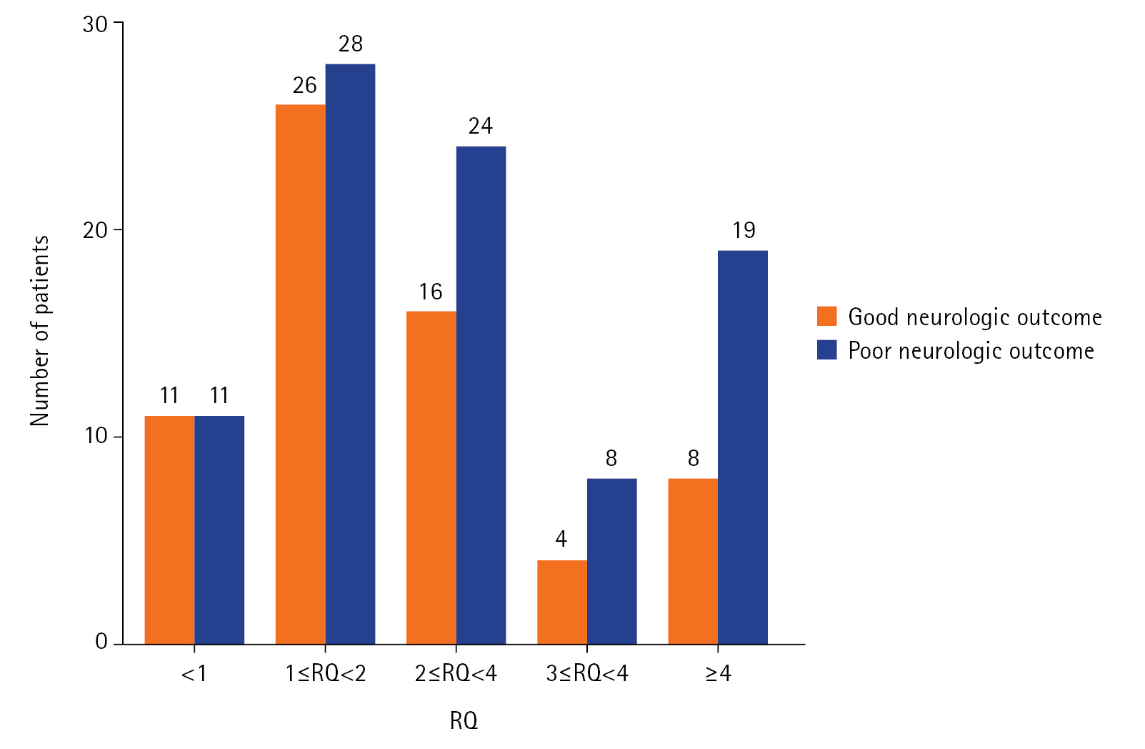
The distribution of patients according to the respiratory quotient. The number of patients according to the respiratory quotient (RQ).

Initial tissue hypoxia parameters and clinical outcome
The predictive power of serum lactate level, RQ, and ΔpCO2 for poor neurologic outcome is shown in Figure 3. The AUROC of serum lactate level was 0.635 (95% CI, 0.542–0.727; P<0.001), and the cut-off point was 7.1 (sensitivity and specificity were 60.0 and 66.2, respectively). The AUROC of RQ was 0.609 (95% CI, 0.519–0.699; P<0.001) and the cut-off point was 1.7 (sensitivity and specificity were 72.2 and 49.2, respectively). Finally, the AUROC of ΔpCO2 was 0.579 (95% CI, 0.487–0.670; P<0.001) and the cut-off point was 11.2 (sensitivity and specificity were 43.3 and 75.4, respectively).
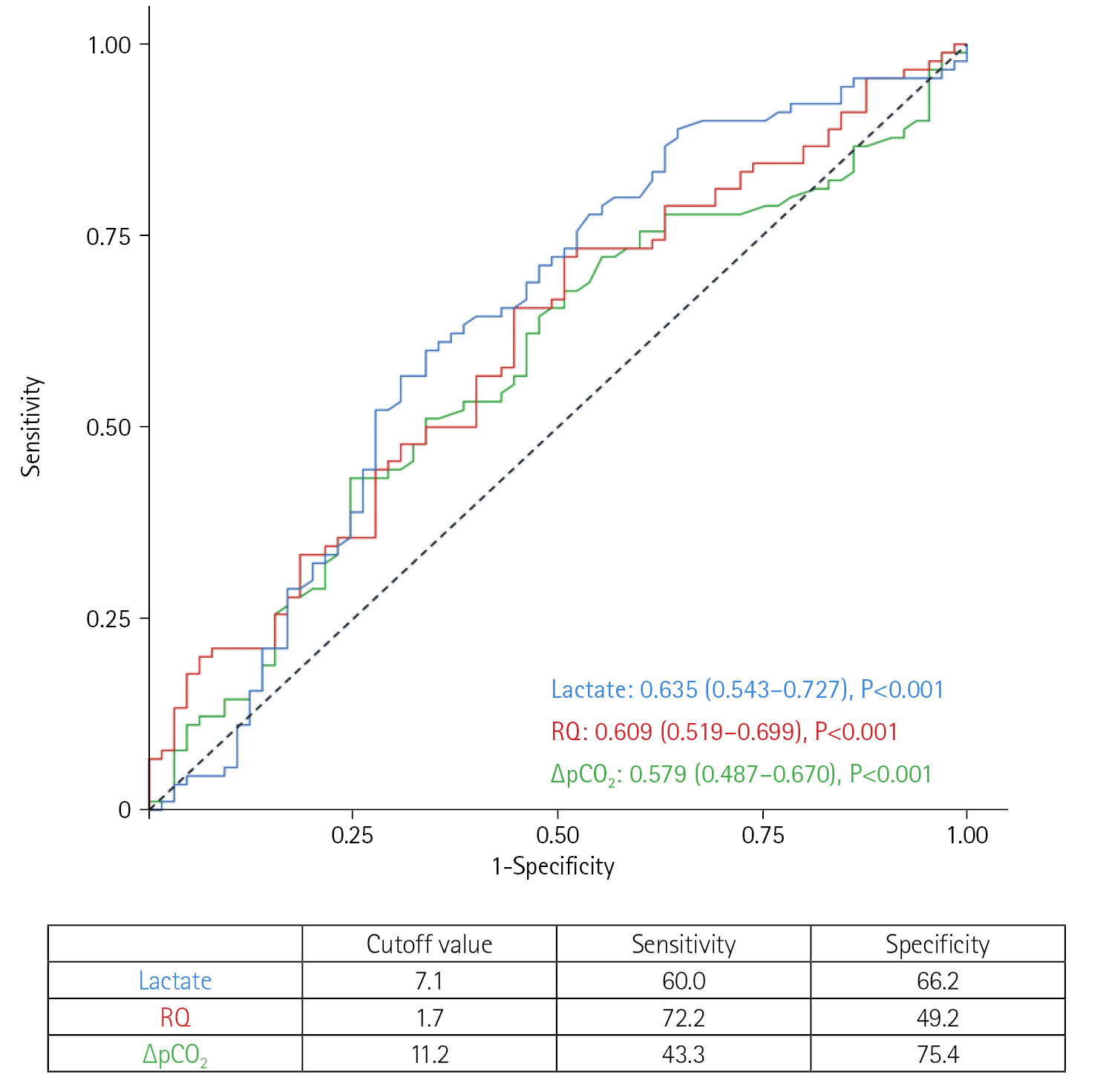
Receiver operating characteristics curves of hypoxia parameters. Values are presented as median (interquartile range). RQ: respiratory quotient; ΔpCO2: venous-arterial difference in CO2 tension.
Predictors for Poor Neurologic Outcome
The results of univariable and multivariable analysis with the logistic regression model for poor neurologic outcome are presented in Table 4. In multivariate analysis, age, CPR to pump-on time, and lactate over 7.1 mmol/L were independently associated with poor neurologic outcome in patients who received ECPR.

Factors associated with poor neurologic outcome
DISCUSSION
The current study investigated the ability of RQ measured within the first 24 hours after ECPR to predict neurologic outcomes in a relatively large number of ECPR cases. The main clinical findings of this study were as follows: (1) The median RQ was 2.0 (IQR, 1.4–3.0) after ECPR, which was almost twice the normal value. (2) Although RQ was significantly higher in patients with poor neurolgoic outcome, it was not independently associated with poor neurologic outcomes in multivariable logistic regression analysis. (3) Among tissue hypoxia markers studied, only serum lactate >7.1 mmol/L was independently associated with poor neurologic outcomes (odds ratio, 2.26; 95% CI, 1.01–5.04; P=0.047).
The clinical significance of RQ has been studied mainly in the context of septic shock. In previous studies involving patients with acute circulatory failure, usually due to septic shock, investigators found that high RQ was associated with absence of improvement in hyperlactemia [10], increase in oxygen consumption after fluid challenge [11,19], and presence of hyperlactemia [16]. In some studies, patients who had both high RQ and hyperlactemia had the worst outcome [20,21]. Finally, higher RQ was also associated indices of microcirculatory failure using near-infrared spectroscopy [22]. These studies suggest that RQ has potential as marker of tissue hypoxia.
In this study, most of the patients had abnormal RQ of >1 which is marked different compared with few small studies performed in post-CPR patients undergoing targetted temperature after ROSC. In this patient population, median RQ values were less than 1.0 [13,23,24]. There are possible explanations for the discrepancy between RQ in patients who received conventional CPR and our ECPR cohort. First, all patients included in studies of RQ after conventional CPR underwent TTM. Hypothermia may diminish the metabolic rate and thus oxygen consumption and carbon dioxide production, which may have influenced RQ values. However, the use of TTM did not affect RQ in this study. RQ was 2.1 (IQR, 1.6–3.4) in patients who received TTM (n=33) and 2.0 (IQR, 1.3–2.9) in patients who did not (n=122) (P=0.493). Second, the timing of blood sampling to calculate RQ was different among studies, which may have influenced the results. Holzinger et al. [13] measured O2 consumption and CO2 production 12 to 24 hours after the patient reached the target temperature of 33 ℃. In the study by Uber et al. [23], blood samples were drawn 5.0 to 18.7 hours (median, 9.9 hours) after the ROSC. Third, there may have been selection bias that could have influenced the results. For example, in this study, patients who died within the first 24 hours because of refractory shock were excluded because we wanted to evaluate the clinical significance of tissue hypoxia parameters in patients with the potential to benefit from hemodynamic optimization. Had these patients not been excluded, the level of RQ might have been different. In addition, each study of RQ in conventional CPR patients had different inclusion and exclusion criteria, so the study populations differed slightly.
The prognostic value of serum lactate level in patients after conventional CPR has been reported in previous studies [6,25-27]. Furthermore, lactate is known to be associated with poor clinical outcomes in patients who receive ECMO [28]. Usually, the in vivo production and consumption of lactate balance each other, so that serum lactate levels are constant. However, in local or general tissue hypoxia, impaired mitochondrial oxidation leads to overproduction and underutilization of lactate. Furthermore, combined metabolic acidosis contributes to decreased hepatic removal of lactate [29]. Thus, lactate has been used as a valuable clinical marker to guide resuscitation. It is unclear as to why RQ was inferior to lactate in predicting poor neurologic outcomes in patients who received ECPR in this study. One possibility is that blood gas measurements may be influenced by sweep gas and ECMO flow rate, which clinicians could calculate. In their recent study on the theoretical effect of extracorporeal CO2 removal on native lung RQ, Cipriani et al. suggested that native lung RQ increases when more oxygen is delivered extracorporeally [30]. Although veno-arterial ECMO and extracorporeal CO2 removal differ in many ways, this may be a clue to explaining the poor utility of RQ as a marker of tissue hypoxia in patients who receive ECPR. In other words, not only tissue hypoxia but also the amount of oxygen delivered by ECMO might have affected RQ in patients who received ECPR. The effect of sweep gas and ECMO flow on RQ needs further investigation. Second, we used the difference in CO2 partial pressure as a surrogate for the difference in CO2 content to calculate RQ. Although there is good correlation between these two values, these two values are not identical, especially when the CO2 partial pressure is not in the physiological range [11,20,31]. Clinically, calculating serum CO2 content requires complex equations, making them impractical to be used at the bedside. Therefore, in many stuides, the CO2 partial pressure is used as a substitute for the CO2 content in the calculation of the RQ [17,21,22,32]. Third, the exact proportion of ECMO flow compared with native flow in the radial artery could not be measured, which may have influenced blood gas measurements. The proportion of ECMO flow might have increased when native cardiac function decreased. Fourth, the diet might have affected patients' metabolism and thus the RQ level. Li et al. [33] showed an significant increase in RQ from 0.6 to 1.0 with increasing caloric intake in five pediatric patients receiving ECMO.
There were some limitations to our study. First, this study was conducted with data collected over a long period. During this time, post-arrest management has changed, which could have affected the results. Second, selection bias might have affected the results because this was a retrospective observational study conducted in a single center. In our study, although patients that were excluded due to insufficient data to calculate RQ had similar neurologic outcomes to patients included in this study, some of the outcomes were different between the two groups (data not shown). These differences in different patient groups could have affected the result of this study. Third, to calculate RQ, arterial and venous blood samples drawn within 30 minutes were used in this study. It is not known how this small difference in sampling times could have influenced the overall result. Fourth, more accurate method of measuring RQ such as indirect calorimetry was not used in this study. Future studies using this method may prove to be useful. Finally, although we used a data from a relatively large cohort of ECPR patients, the statistical power might not have been enough. Larger prospective studies are needed to evaluate the utility of RQ as a predictor of poor neurologic outcomes. In patients who received ECPR, RQ was not independently associated with poor neurologic outcome. Further studies on this subject is needed.
KEY MESSAGES
▪ The median respiratory quotient (RQ) measured within 24 hours of extracorporeal cardiopulmonary resuscitation (ECPR) was 2.0 (interquartile range, 1.4–3.0), which was almost twice the normal value. The RQ was not associated with poor neurologic outcomes in patients who received ECPR.
▪ However, lactate levels above 7.1 mmol/L was associated with poor neurologic outcomes along with other variables such as age, and cardiopulmonary resuscitation to extracorporeal membrane oxygenation pump-on time.
Notes
CONFLICT OF INTEREST
No potential conflict of interest relevant to this article was reported.
FUNDING
This work was supported by the Korea Medical Device Development Fund grant funded by the Korea government (the Ministry of Science and ICT, the Ministry of Trade, Industry and Energy, the Ministry of Health & Welfare, the Ministry of Food and Drug Safety) (project no. 1711138313, KMDF_PR_20200901_0159).
AUTHOR CONTRIBUTIONS
Conceptualization: YIL, JHY, GYS. Data curation: YIL, REK. Formal analysis: YIL, REK, GYS. Funding acquisition: GYS. Methodology: JHY, CRC, GYS. Project administration: JHY, CRC, GYS. Visualization: YIL, REK. Writing–original draft: YIL, REK, GYS. Writing–review & editing: SJN, JAR, YHC.
Acknowledgements
This work was revised from 2022 Master's thesis of Yun Im Lee, Sungkyunkwan University School of Medicine.