Abstract
- Systemic tumor embolisms after pulmonary resections for malignancy are rare, but usually severe and sometimes fatal.
Here, we report a case of a 70-year-old woman who underwent pulmonary resection for lung cancer and subsequently developed acute arterial occlusion of the lower extremities caused by a tumorous embolus.
-
Keywords: embolectomy; embolism; femoral artery; lung neoplasms
Embolism of peripheral arteries originating from malignant tumors is considered a rare manifestation of cancer.[1] Although uncommon, during the pulmonary resection, a fragment of tumor that has invaded a pulmonary vein can embolize and result in arterial occlusion.[2] Symptoms are related to the location of emboli and the most common events are lower extremity, cerebral, myocardial, and limb ischemic events. Here, we present the case of a 70-year-old woman with tumor embolism of the both lower extremities after left pneumonectomy for lung cancer, which was treated successfully with surgical intervention.
Case Report
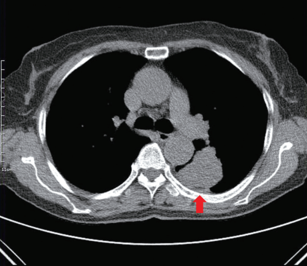
A 70-year-old female, with no previous medical history, was diagnosed centrally located 5.1x3.5 cm-sized mass on left lower lung through medical healthcare check-up. Bronchoscopy demonstrated mass involvement on left distal main bronchus from left lower lobar bronchus. Biopsy and cytologic examination were performed, and the results were positive for sarcomatoid carcinoma. A computerized tomography of chest showed a pulmonary mass in the left lower lung but no distant metastasis was observed (Fig. 1). Left pneumonectomy was performed without any intra-operative events and invasion of the ipsilateral pulmonary vein was identified during the procedure. Immediately after admission to the surgical intensive care unit (ICU) for postoperative management, the patient complained of numbness and pain below right ankle. On physical examination, the right lower extremity was cold with pallor noted on the plantar surface of the right foot. The patient’s dorsalis pedis pulse was non-palpable providing evidence of acute arterial occlusion. Promptly, a handheld vascular Doppler evaluation was performed and absent flow in the right dorsalis pedis artery was demonstrated. Bedside ultrasound of both lower extremities was emergently performed. Hyperechoic material in the right superficial femoral artery extending to the popliteal artery was confirmed (Fig. 2). Color flow Doppler mode of the same vessels revealed no flow below popliteal artery. To identify either acute or chronic arterial occlusion, contrast-enhanced computed tomography angiography was performed and was consistent with arterial occlusion on the same site by acute thromboembolism (Fig. 3A). Emergency embolectomy utilizing No. 3 and 4 Fogarty catheters was performed 6 hours after lung cancer surgery. Fleshy long thromboembolic materials involving the superficial femoral artery and popliteal artery were extracted and the patient had complete return of blood flow and pulse to the lower extremity. To prevent compartment syndrome by revascularization, additional mini-fasciotomy on the right lower extremity was performed. Nevertheless, the day after embolectomy, the patient showed signs of revascularization syndrome that responded favorably to medical treatment including intravenous hydration and bicarbonate. In addition, the patient received intravenous heparin followed by oral anticoagulant therapy. The thromboembolic specimens were confirmed pathologically as tumors of the same type as the primary pulmonary tumor, a sarcomatoid carcinoma. The pathologic TNM stage of primary pulmonary tumor was T2bN1M0. The patient’s recovery was uneventful and discharged on the 22nd postoperative day (Fig. 3B).
Discussion
Non-traumatic acute peripheral arterial occlusion is most commonly caused by thrombosis (85%) followed by embolism (15%).[3] Embolism of the peripheral arteries originating from malignant tumors is considered an infrequent manifestation of cancer. Tumor fragments are responsible for small percentage of peripheral emboli. Tumors of the heart are most often implicated and atrial myxomas are the most common of these, reported in 30% of series.[4] Although uncommon, lung cancer should be considered a possible source of emboli of the extremities, particularly when neoplastic lesions have invaded the pulmonary veins. It is more frequently associated with pneumonectomy than with lesser pulmonary resections. Considering the sites of tumor emboli location, the most frequently involved arteries are those of lower extremities (56.7%) followed by those of the cerebral circulation (34.6%).[5] Symptoms are related to the location of the emboli and most common events are lower extremity, cerebral, myocardial, and limb ischemic events. Patient’s outcome is also related to the site of embolus lodgment and acute embolisms to the coronary, cerebral, and mesenteric arteries usually have a poor prognosis. The overall success rate of embolectomy in lower extremities has been reported to be 84%. The prognosis of patients after successful embolectomy is determined by the TNM staging and their underlying disease. These tumor embolisms cannot be considered distant metastases of the primary tumor, since the vascular endothelium in the area where embolization occurs is not affected.[6]
The first case of arterial tumor embolism was reported in 1904 by Busse[7] in 39-year-old female presented with choriocarcinoma and pulmonary metastases. The majority were associated with primary (44.2%) or secondary lung cancer (31.7%) and primary tumors of aorta (13.4%). The first successful embolectomy for tumor emboli was performed by Groth[8] in 1940 in a patient with tibial sarcoma and pulmonary metastases who, after penumonectomy, developed acute embolism of the left common femoral artery. In 1951, Aylwin[9] suggested that the primary pulmonary veins were ligated as a preventive measure before mobilization of the tumor to avoid this infrequent operative complication. In this case, pulmonary vein involvement of main pulmonary mass was first encountered at the time of surgery. We tried to avoid unnecessary lung manipulation and performed early ligation of the pulmonary vein in order to prevent detachment and subsequent embolization of tumor. Unfortunately, even with precautions, peripheral arterial tumor emboli occurred immediately after transferring to ICU.
How well a patient does depended on the rapid and accurate diagnosis of embolic vessels and prompt surgical intervention, especially within 6 hours after presence of symptoms. Physical examination is important in determining the level of arterial occlusion, as this will assist in determining patient management.[10] The moment the patient developed numbness and pain in lower extremity after admitting ICU, we performed rapid physical examination and assessment. Because the patient who underwent pneumonectomy should be monitored in the ICU, a handheld vascular Doppler evaluation was promptly performed to identify an arterial occlusion. Next, a bedside ultrasound of the both lower extremities was emergently performed. The bedside ultrasound may be a feasible screening test to quickly identify the location of an arterial occlusion.[11] The handheld vascular Doppler and bedside ultrasound could be considered the easiest and safest diagnostic procedure for directly depicting thrombi in the extremities. Moreover, Doppler evaluation of signals of all extremities can identify evidence of contralateral chronic occlusion disease. By the two methods, we confirmed the acute lower peripheral arterial occlusions and the patient was emergently taken to the operating room for arterial embolectomy 6 hours after lung cancer surgery. As a result, she was successfully treated and discharged without complications. Following the diagnosis of critical limb ischemia by embolic occlusions, emergency embolectomy is typically directed toward reversing ischemia and minimizing organ damage, followed by long-term therapy and secondary prevention.
In conclusion, acute peripheral arterial tumorous embolism after lung cancer surgery is a rare cause of acute arterial occlusion but severe and even fatal complication causing limb-threatening ischemia. Early diagnosis and prompt surgical embolectomy will bring the patient good prognosis.
NOTES
-
No potential conflict of interest relevant to this article was reported.
Fig. 1.Chest computed tomography scan showing a 5.1×3.5 cm pulmonary mass (arrow) in the left lower lobe.

Fig. 2.Echogenic thrombus filling (arrow) in the right superficial artery on transverse B-mode ultrasonography.

Fig. 3.Acute thrombotic occlusion (arrow) in the right mid-superficial femoral artery to proximal popliteal artery on preoperative computed tomography angiography (A) and revascularization of the same vessels on postoperative computed tomography angiography (B).

References
- 1. Morsey H, Aslam M, Standfield N. Tumor embolization causing acute ischemia with sometimes fatal results. Case report and review of literature. Int Angiol 2004;23:82-4.PubMed
- 2. Whyte RI, Starkey TD, Orringer MB. Tumor emboli from lung neoplasms involving the pulmonary vein. J Thorac Cardiovasc Surg 1992;104:421-5.ArticlePubMed
- 3. Ouriel K, Veith FJ, Sasahara AA. A comparison of recombinant urokinase with vascular surgery as initial treatment for acute arterial occlusion of the legs. Thrombolysis or Peripheral Arterial Surgery (TOPAS) Investigators. N Engl J Med 1998;338:1105-11.ArticlePubMed
- 4. Tsao JH, Lo HC, How CK, Yen DH, Huang CI. Embolic occlusion of the aorta caused by cardiac myxoma. Resuscitation 2010;81:511. ArticlePubMed
- 5. Xiromeritis N, Klonaris C, Papas S, Valsamis M, Bastounis E. Recurrent peripheral arterial embolism from pulmonary cancer. Case report and review of the literature. Int Angiol 2000;19:79-83.PubMed
- 6. Marinos T, Bitzikas G, Lymperiadis D, Galanos O. Peripheral tumor emboli to both lower extremity arteries after pneumonectomy for extensive pulmonary cancer. J Cardiovasc Surg (Torino) 2004;45:592-3.PubMed
- 7. Busse A. Ueber sarkomatose entartung der myone. Dtsch Med Wochenschr 1904;30:373. Article
- 8. Groth KE. Tumor embolism of the common femoral artery, treated by embolectomy and heparin. Surgery 1940;8:617-32.
- 9. Aylwin JA. Avoidable vascular spread in resection for bronchial carcinoma. Thorax 1951;6:250-67.ArticlePubMedPMC
- 10. O’Connell JB, Quiñones-Baldrich WJ. Proper evaluation and management of acute embolic versus thrombotic limb ischemia. Semin Vasc Surg 2009;22:10-6.ArticlePubMed
- 11. Rolston DM, Saul T, Wong T, Lewiss RE. Bedside ultrasound diagnosis of acute embolic femoral artery occlusion. J Emerg Med 2013;45:897-900.ArticlePubMed
Citations
Citations to this article as recorded by


 PubReader
 ePub Link
 Cite
Cite