Articles
- Page Path
- HOME > Acute Crit Care > Volume 37(3); 2022 > Article
-
Image in critical care
Neurology Contrast media mimicking subarachnoid hemorrhage after intrathecal injection in a patient with Creutzfeldt-Jakob disease -
Taegyun Kim1,2

-
Acute and Critical Care 2022;37(3):474-476.
DOI: https://doi.org/10.4266/acc.2022.00339
Published online: July 4, 2022
1Department of Emergency Medicine, Seoul National University Hospital, Seoul, Korea
2Department of Emergency Medicine, Seoul National University College of Medicine, Seoul, Korea
- Corresponding author: Taegyun Kim Department of Emergency Medicine, Seoul National University Hospital, 101 Daehak-ro, Jongno-gu, Seoul 03080, Korea Tel: +82-2-2072-3257, Fax: +82-2-741-7855, E-mail: kimtagyun@snuh.org
• Received: March 15, 2022 • Revised: April 28, 2022 • Accepted: April 30, 2022
Copyright © 2022 The Korean Society of Critical Care Medicine
This is an Open Access article distributed under the terms of the Creative Commons Attribution Non-Commercial License (http://creativecommons.org/licenses/by-nc/4.0/) which permits unrestricted non-commercial use, distribution, and reproduction in any medium, provided the original work is properly cited.
- 1,409 Views
- 141 Download
- 1 Web of Science
This article has been corrected. See "Contrast media mimicking subarachnoid hemorrhage after intrathecal injection in a patient with Creutzfeldt-Jakob disease" in Volume 37 on page 693.
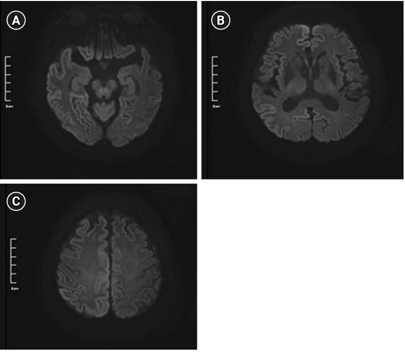
Figure 1. Brain diffusion-weighted imaging at b1000 at the levels of (A) midbrain, (B) thalamus, and (C) frontal and occipital lobes. Multifocal diffusion restriction can be seen along the bilateral cerebral cortex, especially prominent on the right parietal area.

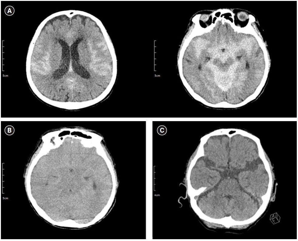
Figure 2.Serial brain computed tomography images. (A) At midnight on the day of fluoroscopy-guided cerebrospinal fluid drainage, diffuse high attenuation was seen in the subarachnoid space (66 HU). (B) Twelve hours after initial observation, diffuse sulcal high attenuation was still noted (59 HU); however, it was more dispersed into the subarachnoid space. (C) After 24 hours, high attenuation had nearly disappeared (21 HU). HU: hounsfield unit.

- 1. Hasan TF, Duarte W, Akinduro OO, Goldstein ED, Hurst R, Haranhalli N, et al. Nonaneurysmal "pseudo-subarachnoid hemorrhage" computed tomography patterns: challenges in an acute decision-making heuristics. J Stroke Cerebrovasc Dis 2018;27:2319-26.ArticlePubMed
- 2. Oh CH, An SD, Choi SH, Ji GY. Contrast mimicking a subarachnoid hemorrhage after lumbar percutaneous epidural neuroplasty: a case report. J Med Case Rep 2013;7:88. ArticlePubMedPMCPDF
- 3. Platt A, Ammar FE, Collins J, Ramos E, Goldenberg FD. Pseudo-subarachnoid hemorrhage and gadolinium encephalopathy following lumbar epidural steroid injection. Radiol Case Rep 2020;15:1935-8.ArticlePubMedPMC
- 4. Sasaki Y, Ishii K, Ishii I. Myelography-associated pseudo-subarachnoid hemorrhage. Vis J Emerg Med 2016;5:25-6.Article
References
Figure & Data
References
Citations
Citations to this article as recorded by 
 PubReader
 ePub Link
 Cite
Cite