Abstract
-
Background
- The external jugular vein (EJV) is a useful intravenous (IV) cannulation site for anesthesiologists, although it has a relatively high failure rate. Unlike other central veins, visualization of the EJV is important during IV cannulation, and the Valsalva maneuver distends the jugular venous system. However, the relationship between the maneuver and EJV visibility remains unknown. This study compared EJV visibility before and after the Valsalva maneuver.
-
Methods
- This was a prospective observational study that included 200 participants. After the induction of anesthesia, EJV visibility grade, depth from the skin to the EJV superficial surface (EJV depth), and EJV cross-sectional area (CSA) before the Valsalva maneuver were measured. The same parameters were measured after the Valsalva maneuver was performed. The EJV visibility grade was defined as grade A: good appearance and good palpation, grade B: poor appearance and good palpation, and grade C: poor appearance and poor palpation.
-
Results
- Patient body mass index and EJV depth affected the EJV visibility grade before the Valsalva maneuver (p < 0.05), although EJV CSA did not. The Valsalva maneuver distended EJV CSA and reduced EJV depth, although these changes were not correlated with EJV visibility grade. With regard to EJV visibility, 34.0% of grade B cases and 20.0% of grade C cases were improved by the Valsalva maneuver.
-
Conclusions
- Although the Valsalva maneuver improved EJV CSA and EJV depth, it did not greatly affect EJV visibility grade.
-
Keywords: jugular veins; ultrasonography; Valsalva maneuver
Introduction
The external jugular vein (EJV) is a commonly used route for intravenous (IV) cannulation during general anesthesia. EJV cannulation is also indicated in the intensive care unit when massive and/or quick fluid administration or central venous pressure measurements are needed.[1,2] The EJV has many benefits compared to other IV routes, including the central veins, as it is easy to visualize (even during cardiac arrest), is located in a superficial location, and is relatively easy to access without imaging modalities.[1,3] However, the EJV route also has a relatively low success rate.[4] According to a few reports, the low success rate of the EJV route is related to its poor visibility and anatomical variability, as well as the practitioner’s technique.[5,6] To increase the success rate of EJV cannulation, many methods have been described, including distending the jugular venous system[7] and using ultrasound to visualize the EJV.[8-10] In addition, the Valsalva maneuver, Trendelenburg’s position, and abdominal compression can dilate the EJV and provide a similar beneficial effect.[7,11] However, the primary outcomes in the previous studies were not the EJV’s visibility, but rather the cross-sectional area (CSA) as measured via ultrasound.[12] Moreover, the factors related to the improved EJV visibility are not yet known.
We designed this prospective observational study to evaluate the hypothesis that distension of the EJV CSA using the Valsalva maneuver would improve the EJV visibility. Therefore, the study’s primary objective was to compare the EJV visibility before and after the Valsalva maneuver in patients who were under general anesthesia. The secondary objectives were to compare the EJV CSA and depth from the skin to the superficial surface of the EJV (EJV depth) using ultrasonography before and after the Valsalva maneuver, and to evaluate the relationship between changes in EJV visibility grade and other variables.
Materials and Methods
This prospective observational study was conducted between March 1, 2012 and August 31, 2012 in the operating room of a university hospital. The institutional review board approved the study’s protocol, and all patients provided their written informed consent to participate in this study.
Two hundred participants were enrolled, and their medical history was taken at their enrollment. The inclusion criteria were patients who had an American Society of Anesthesiologists physical status classification of 1 or 2 and who were > 20 years old. The exclusion criteria included an the presence of an abdominal mass, ascites, pregnancy, increased intracranial pressure, lung disease, previous neck surgery (cervical spine, thyroid, and ear, neck, or throat surgery), neck mass, or perioperative hypotension.
Several variables such as sex, age, body mass index (BMI), EJV depth (Fig. 1A), and EJV CSA (Fig. 1B) were evaluated for their relationship with EJV visibility. EJV visibility was defined as grade A: good appearance and good palpation, grade B: poor appearance and good palpation, and grade C: poor appearance and poor palpation.
All patients were placed under general anesthesia, which was induced by propofol at 2 mg/kg, followed by tracheal intubation after intravenous administration of rocuronium at 0.6 mg/kg. Anesthesia was maintained by administration of 6-8 vol% desflurane and a fresh gas flow of 50% oxygen and air at 4 L/min. When the patient’s hemodynamic values had stabilized after the induction of anesthesia, we evaluated the EJV visibility grade, EJV depth, and EJV CSA before and after the Valsalva maneuver using a portable ultrasound system (ACUSON X300, Siemens Medical Solutions, Malvern, PA, USA) with a 5-13 MHz linear transducer. The Valsalva maneuver in the pressure controlled-mode at a plateau airway pressure of 20 cmH2O was performed for 10 s.[13] The EJV visibility grade was evaluated by a single observer to reduce bias. The EJV CSA and EJV depth were measured three times at the cricothyroid membrane level, and the average of these values was used for the analysis. A thick layer of ultrasound gel was applied to prevent EJV compression.
The patient’s blood pressure, heart rate, and airway pressure were recorded during the Valsalva maneuver to monitor for complications. In this clinical trial, we defined hypotension and bradycardia as a 20% reduction in the initial value. If hypotension or bradycardia occurred during the Valsalva maneuver, the procedure was stopped and the hypotension was managed by changing the patient’s position and using an appropriate vasopressor, such as ephedrine.
All data are presented as mean ± standard deviation or number of patients (%). Demographics and baseline variables before the Valsalva maneuver were analyzed using the chi-square test or one-way analysis of variance with a post-hoc least significant difference test, as appropriate. Differences in the EJV visibility, EJV CSA, and EJV depth before and after the Valsalva maneuver were evaluated using paired t-tests. Pearson’s correlation was used to evaluate the relationship between EJV depth before the Valsalva maneuver and other variables such as EJV CSA, BMI, and age. A simple logistic regression analysis was used to evaluate the relationship between improvements in the EJV visibility grade and the other variables. No missing patient data were discovered in this study. A p value of < 0.05 was considered statistically significant, and all statistical analyses were performed using SAS for Windows (version 9.2, SAS Institute Inc., Cary, NC, USA).
Results
Two hundred individuals were recruited for this study and complete data were collected for all participants (Fig. 2). The participants (115 women and 85 men) had an average age of 50 ± 17 years, average weight of 65.1 ± 38.8 kg, and an average height of 160.0 ± 10.0 cm. The demographic and baseline data according to EJV visibility grade classification are presented in Table 1. BMI and the EJV depth before the Valsalva maneuver affected the EJV visibility grade before the Valsalva maneuver, although the EJV CSA was not related to the EJV visibility grade. In addition, a positive correlation was observed between BMI and the EJV depth before the Valsalva maneuver(r = 0.27, p < 0.001).
The Valsalva maneuver significantly affected both the EJV CSA (Fig. 3A) and the EJV depth (Fig. 3B). After the Valsalva maneuver, the EJV CSA increased from 0.16 ± 0.17 cm2 to 0.23 ± 0.19 cm2 (35.6%), and the EJV depth decreased from 2.74 ± 1.18 mm to 2.42 ± 1.06 mm (10.7%). However, no significant changes were observed in the EJV visibility grade, as 34.0% of cases with grade B visibility and 20% of cases with grade C visibility exhibited an improved gross appearance after the Valsalva maneuver (Table 2). To analyze the factors that were associated with improvements in the EJV visibility grade after the Valsalva maneuver, we reviewed several variables, including age, BMI, EJV CSA before the Valsalva maneuver, changes in the EJV CSA due to the Valsalva maneuver, EJV depth before the Valsalva maneuver, and changes in the EJV depth due to the Valsalva maneuver. However, none of these factors were significantly associated with the EJV visibility grade (Table 3). During the Valsalva maneuver, no complications were observed, including hypotension and bradycardia.
Discussion
After the Valsalva maneuver, the EJV CSA and EJV depth changed significantly (Fig. 3), although only 27.2% of the patients (visibility grade B and C) exhibited an improved visibility grade (Table 2). We were unable to identify any factors that were related to the change in EJV visibility grade (Table 3). In addition, even a significant change in the EJV depth after the Valsalva maneuver did not affect the EJV visibility grade, unlike the initial EJV visibility grade (Table 1). We propose two possible explanations for these findings. First, the Valsalva maneuver that we performed may not have been sufficiently forceful to observe an improved EJV visibility grade. Second, unidentified patient factors, such as skin texture, thickness, or color, might be important, although we did not evaluate these factors in this study.
Central veins, including the internal jugular vein, subclavian vein, and femoral vein, are deep structures that are not visible. Therefore they can be cannulated using an anatomical landmark. However, the EJV is a superficial structure that is readily visible, and this visibility is an important factor in the successful cannulation of the EJV, unlike the other central veins.[2] There have been many studies to assess factors associated with success rate of central vein cannulation, including the EJV, and methods to increase the CSA of the central vein.[7,12,14-18] The Valsalva maneuver, abdominal compression, and the Trendelenburg position are common methods for improving venous distension. The Valsalva maneuver has been shown to increase the EJV CSA by 136 ± 23% (95% confidence interval, 121.3-147.5%), and the Trendelenburg position has been shown to increase the EJV CSA by 137 ± 32% (95% confidence interval, 120.7-156.9%).[7] These results are similar to ours, but EJV visibility was not considered in the previous study.
The Valsalva maneuver is performed using a forceful attempted exhalation against a closed glottis, leading to incremental increases in intrathoracic pressure and size of the extrathoracic veins, including the EJV.[14,19] Cardiovascular responses such as heart rate, blood pressure, stroke volume, and venous return, differ with various strain pressures in the Valsalva maneuver.[19] In this clinical trial, we successfully applied a low strain pressure and short duration for the Valsalva maneuver to avoid any cardiovascular complications. [13,19]
Several limitations of this study should be considered when interpreting the results. First, actual cannulation of the EJV was not performed in this study, and we only evaluated various factors that might have been linked to the EJV’s gross appearance. Second, although the EJV depth was associated with the visibility grade before the Valsalva maneuver (Table 1), and subsequently decreased after the Valsalva maneuver (Fig. 3B), the actual change in the visibility grade was minimal (Table 2). Therefore, we cannot comment on the magnitude of in the EJV depth that is needed to improve the visibility grade. Third, only one observer evaluated EJV visibility in this study. Although it is good to increase interobserver reliability, this practice could also have caused bias in the intra-observer reliability. Further studies should incorporate EJV cannulation before and after the Valsalva maneuver. In addition, the success rate of EJV cannulation by the visibility grade should also be explored.
In conclusion, although the Valsalva maneuver increased the EJV CSA and decreased the EJV depth, it did not greatly improve the EJV’s visibility.
NOTES
-
No potential conflict of interest relevant to this article was reported.
Fig. 1.External jugular vein cross-sectional area (A) and external jugular vein depth measured by ultrasonography (B). D1: depth of 1; C: circumference.
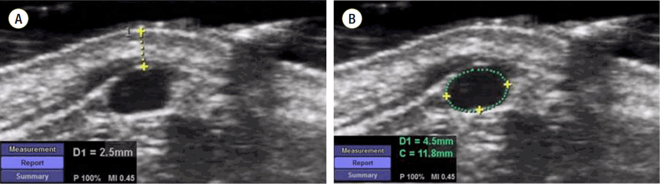
Fig. 2.Flow diagram of the patients.
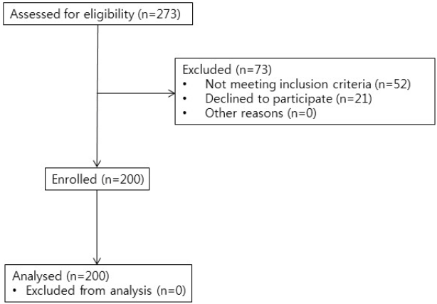
Fig. 3.Comparisons of external jugular vein cross-sectional area (A) and external jugular vein depth before and after the Valsalva maneuver at each EJV visibility grade (B). Visibility grade A: good appearance and good palpation of the EJV; visibility grade B: poor appearance and good palpation of the EJV; visibility grade C: poor appearance and poor palpation of the EJV. The blue-colored box represents EJV CSA or depth before the Valsalva maneuver, and the green-colored box represents EJV CSA or depth after the Valsalva maneuver. The box plot shows the median (solid line), interquartile ranges (box), and values within 1.5 interquartile ranges from each side of the box (whiskers). Outliers are indicated by solid circles. EJV: external jugular vein; CSA: cross-sectional area of the external jugular vein; EJV depth: depth from the skin to the external jugular vein superficial surface; *p < 0.05 compared with EJV visibility before the Valsalva maneuver.

Table 1.Demographics and baseline variables before performance of the Valsalva maneuver
|
Visibility grade A (n = 97) |
Visibility grade B (n = 53) |
Visibility grade C (n = 50) |
p-value |
|
Sex (M/F) |
45/52 |
22/31 |
17/43 |
0.352 |
|
Age (years) |
52 ± 18 |
45 ± 15 |
50 ± 17 |
0.081 |
|
BMI (kg/m2) |
22.80 ± 3.61 |
24.12 ± 3.13*
|
24.14 ± 3.39*
|
0.025 |
|
EJV CSA (cm2) |
0.19 ± 0.17 |
0.18 ± 0.16 |
0.14 ± 0.17 |
0.159 |
|
EJV depth (mm) |
2.31 ± 1.04 |
2.98 ± 1.12*
|
3.30 ± 1.24*
|
< 0.001 |
|
MBP (mmHg) |
70.08 ± 6.87 |
71.19 ± 7.05 |
69.62 ± 8.05 |
0.517 |
|
HR (beats/min) |
72.88 ± 11.15 |
73.84 ± 11.05 |
73.09 ± 11.33 |
0.896 |
Table 2.External jugular vein visibility grade before and after the Valsalva maneuver
|
Visibility grade |
|
Before the Valsalva maneuver |
|
A |
|
|
B |
|
|
C |
|
|
Number |
|
97 |
|
|
53 |
|
|
50 |
|
|
After the Valsalva maneuver |
A |
B |
C |
A |
B |
C |
A |
B |
C |
|
Number (%) |
97 (100.0) |
0 (0.0) |
0 (0.0) |
18 (34.0) |
34 (64.1) |
1 (1.9) |
1 (2.0) |
9 (18.0) |
40 (80.0) |
Table 3.Variables associated with improvements in external jugular vein visibility grade
|
Predictor variables |
Odds ratio |
95% confidence interval |
p-value |
|
Age |
1.01 |
0.99 - 1.03 |
0.398 |
|
BMI |
0.94 |
0.84 - 1.05 |
0.246 |
|
EJV CSA before the Valsalva maneuver |
0.54 |
0.07 - 4.08 |
0.549 |
|
% changes in EJV CSA by Valsalva |
1.01 |
1.00 - 1.02 |
0.109 |
|
EJV depth before the Valsalva maneuver |
0.90 |
0.65 - 1.25 |
0.543 |
|
Changes in EJV depth by Valsalva |
0.99 |
0.97 - 1.02 |
0.589 |
References
- 1. Uvelin A, Kolak R, Putnik D. External jugular cannulation is irreplaceable in many situations. Resuscitation 2010;81:773. author reply 774.ArticlePubMed
- 2. Dailey RH. External jugular vein cannulation and its use for CVP monitoring. J Emerg Med 1988;6:133-5.ArticlePubMed
- 3. Pettit J. External jugular cannulation in infants and children. J Infus Nurs 2009;32:93-7.ArticlePubMed
- 4. Lahtinen P, Musialowicz T, Hyppölä H, Kiviniemi V, Kurola J. Is external jugular vein cannulation feasible in emergency care? A randomised study in open heart surgery patients. Resuscitation 2009;80:1361-4.ArticlePubMed
- 5. Costantino TG, Kirtz JF, Satz WA. Ultrasound-guided peripheral venous access vs. the external jugular vein as the initial approach to the patient with difficult vascular access. J Emerg Med 2010;39:462-7.ArticlePubMed
- 6. Giesy J. External jugular vein access to central venous system. JAMA 1972;219:1216-7.Article
- 7. Lewin MR, Stein J, Wang R, Lee MM, Kernberg M, Boukhman M, et al. Humming is as effective as Valsalva’s maneuver and Trendelenburg’s position for ultrasonographic visualization of the jugular venous system and common femoral veins. Ann Emerg Med 2007;50:73-7.ArticlePubMed
- 8. Galinski M, Catineau J, Tazarourte K, Dardel N, Bertrand P, Adnet F, et al. Comparison of historical anatomic landmarks vs. ultrasound guidance for the selection of a needle insertion site for jugular central venous access. Resuscitation 2012;83:e113-4.ArticlePubMed
- 9. Mitre CI, Golea A, Acalovschi I, Mocan T, Caea AM, Ruţă C, et al. Ultrasound-guided external jugular vein cannulation for central venous access by inexperienced trainees. Eur J Anaesthesiol 2010;27:300-3.ArticlePubMed
- 10. Gann M Jr, Sardi A. Improved results using ultrasound guidance for central venous access. Am Surg 2003;69:1104-7.ArticlePubMedPDF
- 11. Scheller MS, Saidman LJ. An aid to identifying the external jugular vein. Anesthesiology 1982;57:546-7.ArticlePubMed
- 12. Bellazzini MA, Rankin PM, Gangnon RE, Bjoernsen LP. Ultrasound validation of maneuvers to increase internal jugular vein cross-sectional area and decrease compressibility. Am J Emerg Med 2009;27:454-9.ArticlePubMed
- 13. Zhou Q, Xiao W, An E, Zhou H, Yan M. Effects of four different positive airway pressures on right internal jugular vein catheterisation. Eur J Anaesthesiol 2012;29:223-8.ArticlePubMed
- 14. Verghese ST, Nath A, Zenger D, Patel RI, Kaplan RF, Patel KM. The effects of the simulated Valsalva maneuver, liver compression, and/or Trendelenburg position on the cross-sectional area of the internal jugular vein in infants and young children. Anesth Analg 2002;94:250-4.ArticlePubMed
- 15. Beddy P, Geoghegan T, Ramesh N, Buckley O, O’Brien J, Colville J, et al. Valsalva and gravitational variability of the internal jugular vein and common femoral vein: ultrasound assessment. Eur J Radiol 2006;58:307-9.ArticlePubMed
- 16. Yuki K, Chilson K, Odegard KC, DiNardo JA. Trendelenburg position, simulated Valsalva maneuver, and liver compression do not alter the size of the right internal jugular vein in patients with a bidirectional Glenn shunt. Anesth Analg 2007;105:365-8.ArticlePubMed
- 17. Rippey JC, Pascu O, Jacobs I. Abdominal compression effectively increases the size of the common femoral vein, as measured by ultrasonography. Ann Emerg Med 2008;52:446-52.ArticlePubMed
- 18. Kim JT, Park CS, Kim HJ, Lee JM, Kim HS, Kim CS, et al. The effect of inguinal compression, Valsalva maneuver, and reverse Trendelenburg position on the cross-sectional area of the femoral vein in children. Anesth Analg 2009;108:1493-6.ArticlePubMed
- 19. Looga R. The Valsalva manoeuvre--cardiovascular effects and performance technique: a critical review. Respir Physiol Neurobiol 2005;147:39-49.ArticlePubMed
Citations
Citations to this article as recorded by


 PubReader
 ePub Link
 Cite
Cite


