Damage control strategy in bleeding trauma patients
Article information

Abstract
Hemorrhagic shock is a main cause of death in severe trauma patients. Bleeding trauma patients have coagulopathy on admission, which may even be aggravated by incorrectly directed resuscitation. The damage control strategy is a very urgent and essential aspect of management considering the acute coagulopathy of trauma and the physiological status of bleeding trauma patients. This strategy has gained popularity over the past several years. Patients in extremis cannot withstand prolonged definitive surgical repair. Therefore, an abbreviated operation, referred to as damage control surgery (DCS), is needed. In addition to DCS, the likelihood of survival should be maximized for patients in extremis by providing appropriate critical care, including permissive hypotension, hemostatic resuscitation, minimization of crystalloid use, early use of tranexamic acid, and avoidance of hypothermia and hypocalcemia. This review presents an overview of the evolving strategy of damage control in bleeding trauma patients.
INTRODUCTION
In South Korea, injury is a major cause of death, especially for younger age groups, and according to a 2015 nationwide survey, approximately 30% of deaths due to trauma would have been preventable if adequate treatment had been administered [1]. Furthermore, the top cause of death in trauma is uncontrollable bleeding [2]. “Damage control” is a term used in the maritime industry to refer to the emergency management of situations that may cause a vessel to sink. At the scene of an accident, only temporary, limited, urgent repairs are conducted to prevent sinking and then the damaged ship is brought to the dock for definitive repairs. In a similar sense, damage control surgery (DCS) refers to operations performed in patients whose condition is in extremis due to bleeding. In DCS, many procedures are omitted in order to focus on preserving vital aspects of physiology, based on the concept that severely injured patients cannot withstand the prolonged procedures and physiological insults associated with definitive repair [3-6]. In other words, definitive care of trauma patients in extremis in a single operation is like fully repairing a damaged ship at the scene without adequate equipment.
DAMAGE CONTROL RESUSCITATION
Damage control resuscitation (DCR) is an extension of the concept of damage control in severe trauma patients and accepted as complementary to DCS. Conceptually, DCR encompasses not only DCS, but all critical care approaches that correct trauma-induced coagulopathy and provide optimal resuscitation [7-11]. DCR incorporates permissive hypotension to prevent clots from dislodging, minimization of crystalloid use, hemostatic resuscitation, body rewarming, and early hemorrhage control (Figure 1).
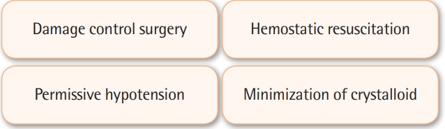
Four main components of damage control resuscitation.
PERMISSIVE HYPOTENSION AND MINIMIZATION OF CRYSTALLOID USE
Permissive hypotension is also known as hypotensive resuscitation. The main concept is restricting the amount of resuscitation fluid and vasopressors to maintain blood pressure in the lower-than-normal range until the bleeding is controlled. Achieving a balance between organ perfusion and hemostasis is crucial for bleeding trauma patients, and clinicians must walk the tightrope between bleeding and hypotensive shock. If the blood pressure is higher, it is more likely that any clots that have formed will be dislodged by the high pressure of the blood and the amount of bleeding will increase. Maintaining normal blood pressure in an uncontrolled bleeding situation may therefore worsen survival, which provides a rationale for maintaining lower-than-normal blood pressure. The target systolic blood pressure of permissive hypotension is 80–90 mm Hg; however, permissive hypotension is only a temporary tool to be used until the source of bleeding is controlled because prolonged hypotension can cause ischemic damage to end-organs, including the brain and kidney, and worsen lactic acidosis [12]. Therefore, in patients with severe traumatic brain injury (TBI), this rule does not apply, and the new target for TBI patients with glasgow coma scale ≤ 8 is a mean arterial pressure > 80 mm Hg [12].
Target of Permissive Hypotension
Another issue is the minimization of crystalloid use. The Advanced Trauma Life Support (ATLS) guidelines traditionally recommended the infusion of 2 L of crystalloid in patients with suspected hemorrhagic shock [13]. However, in the current 10th edition of the ATLS guidelines, only 1 L of crystalloid (including the amount in the pre-hospital setting) is suggested [14]. Excessive crystalloid use is associated with a risk of dilutional coagulopathy because crystalloids contain no blood products (e.g., red blood cells [RBCs] and coagulation factors) and pose a risk of hypothermia, acute lung injury, abdominal compartment syndrome, multi-organ failure, and death [15-17]. A multicenter trial evaluating the minimization of crystalloids for penetrating torso injuries was reported in 2015 [18]. Compared with the standard fluid resuscitation group, the restrictive fluid resuscitation group exhibited lower intraoperative mortality (9% vs. 32%; P < 0.001) and a shorter hospital length of stay (13 vs. 18 days; P = 0.02). In 2017, Harada et al. [19] reported, in an analysis of 10-year trends in crystalloid resuscitation, that the decrease in high-volume crystalloid resuscitation paralleled a reduction of mortality.
HEMOSTATIC RESUSCITATION
Hemostatic resuscitation is now widely accepted as a transfusion therapy for bleeding trauma patients in extremis. The traditional component therapy that only gave packed RBCs (pRBCs) and crystalloids could not replace the coagulation factors consumed by bleeding. The concept of hemostatic resuscitation is the transfusion of blood products (pRBCs, fresh frozen plasma [FFP], and platelets) that closely approximate whole blood. In 2007, Borgman et al. [20] reported a survival benefit for a high ratio of plasma to RBCs in patients who received massive transfusion. A high plasma-to-RBC ratio was independently associated with survival benefits due to reduced death from hemorrhage. Early and aggressive plasma transfusion can reduce mortality from bleeding [20-23].
A high ratio of platelets to RBCs is also very important. In 2008, Holcomb et al. [23] investigated the plasma-to-RBC and platelet-to-RBC ratios in 466 civilian trauma patients who underwent massive transfusions. They divided the patients into four groups according to ratios of FFP to RBCs and platelets to RBCs of 1:2, as follows: (1) high FFP-to-RBC and high platelet-to-RBC ratios, (2) high FFP-to-RBC and low platelet-to-RBC ratios, (3) low FFP-to-RBC and high platelet-to-RBC ratios, and (4) low FFP-to-RBC and low platelet-to-RBC ratios. The patients with high FFP-to-RBC and high platelet-to-RBC ratios had significantly higher survival rates at 6 hours, 24 hours, and 30 days than the other groups.
However, the optimal FFP-to-RBC and platelet-to-RBC ratios remained unknown. In 2015, the Pragmatic, Randomized Optimal Platelet and Plasma Ratios (PROPPR) study was conducted, in which 680 patients were randomized to receive either a 1:1:1 or 1:1:2 ratio of plasma, platelets, and RBCs [24]. Overall mortality was not significantly different between both groups, although death from exsanguination was less common in the 1:1:1 group than in the 1:1:2 group. In conclusion, FFP-to-RBC and platelet-to-RBC ratios of at least 1:2 are recommended for DCR.
BODY REWARMING
Hypothermia is a common problem in the resuscitation of trauma patients because of cold exposure, cold resuscitation fluids, significant blood loss, and shock. The problem is that hypothermia can negatively affect coagulation function. One study showed that relative clotting factor function was sensitively decreased by temperatures ranging from 37°C to 25°C; even if there was no deficiency in clotting factors, their function was severely impaired to less than 10% of normal [25]. In 1994, Gubler et al. [26] demonstrated that dilutional coagulopathy had an additive effect on hypothermia. Hypothermia does not decrease the level of clotting factors or platelets, but directly affects their function. Therefore, treating hypothermia actively with heated fluids, heated blanket, and a warm environment is recommended.
TRANEXAMINIC ACID
Tranexamic acid (TXA) is an anti-fibrinolytic drug that interferes with the binding of plasminogen to fibrin. In theory, TXA can prevent clot breakdown and reduce blood loss in bleeding trauma patients. In 2011, the CRASH-2 trial was conducted to evaluate the effect of early administration of a short course of TXA on death from bleeding trauma [27]. The mortality of the group that received TXA within 1 hour of injury was 5.3%, compared to 7.7% in the placebo group, and TXA did not increase the risk of thrombosis. Patients who received TXA between 1 and 3 hours after the injury also had a survival benefit from bleeding to death. However, if TXA was given more than 3 hours after the injury, the risk of death due to bleeding significantly increased. Therefore, TXA should be first administered within 3 hours from injury. Specifically, 1 g of TXA is administered intravenously or intraosseously, mixed with 100 mL of saline for 10 minutes, followed by another 1 g steady drip over 8 hours.
However, there has been some criticism of the routine use of TXA. In 2016, Moore et al. [28] investigated 2,540 severe trauma patients with an Injury Severity Score >15 and showed that the fibrinolysis shutdown type was the most common, whereas the hyperfibrinolytic type that needed antifibrinolytic agents only accounted for 18% of cases. Therefore, the authors recommended individualization of TXA usage after confirmation of coagulation status through a viscoelastic assessment.
CALCIUM
Calcium is an important cofactor of the coagulation cascade. In bleeding trauma patients who need blood transfusion, hypocalcemia may be a problem because of citrate, a calcium chelating agent contained in many blood products. The effects of hypocalcemia cannot be evaluated through routine laboratory tests, and even 1 unit of citrate-containing blood products can further lower calcium ion levels to the point that they approach critical values of hypocalcemia associated with lower survival [29-33]. Calcium ion levels should be monitored and maintained within the normal range. To correct hypocalcemia, calcium chloride is preferable due to the amount of calcium that it contains, as 10% calcium chloride contains 270 mg of elemental calcium per 10 mL, whereas 10% calcium gluconate contains 90 mg of elemental calcium per 10 mL [12].
DAMAGE CONTROL SURGERY
In the 1980s, an important paradigm shift occurred in the treatment of bleeding trauma patients. DCS is a treatment strategy that focuses on rapid control of hemorrhage and contamination [3,4,34]. Time is very limited for DCS—usually within 90 minutes—because of the concept that severely injured patients cannot withstand the prolonged procedures and physiological insult that would be required for definitive repair. Therefore, the bowel is left in a discontinuous condition and vessels may be kept patent with temporary shunts.
Three steps of DCS
First step is abbreviated operation, including bleeding and contamination control, as well as temporary abdominal closure. Second is critical care for physiological restoration in the intensive care unit. Third is definitive surgery. DCS is now accepted as a component of DCR and is concomitantly conducted with DCR in most cases.
CONCLUSION
The damage control strategy is a very urgent and essential aspect of management for bleeding trauma patients, considering the acute coagulopathy of trauma and these patients’ physiological status. The strategy incorporates an abbreviated operation, referred to as DCS, as well as appropriate critical care including permissive hypotension, hemostatic resuscitation, minimization of crystalloid use, early use of TXA, and avoidance of hypothermia and hypocalcemia. These approaches collectively improve the survival of bleeding trauma patients in extremis.
KEY MESSAGES
▪ Damage control surgery (DCS) focuses on rapid control of hemorrhage and contamination for patients in extremis.
▪ DCS is usually conducted within a very limited time, and non-urgent procedures are omitted or postponed to a subsequent operation.
▪ Damage control resuscitation incorporates a broad range of approaches to save the lives of bleeding trauma patients, including DCS, permissive hypotension, hemostatic resuscitation, minimization of crystalloid use, early use of tranexamic acid, and avoidance of hypothermia and hypocalcemia.
Notes
CONFLICT OF INTEREST No potential conflict of interest relevant to this article was reported.
AUTHOR CONTRIBUTIONS
Conceptualization & Data curation: HC. Formaly analysis & Methodology: MK. Project administration: HC. VIsualization: MK. Writing–original draft: HC. Writing–review & editing: MK.