Sepsis-induced cardiomyopathy is associated with higher mortality rates in patients with sepsis
Article information
 , Anika Sasidharan Nair2, Adarsh Katamreddy2, Jason S Gilbert3, Jee Young You2, Obiageli Lynda Offor2, Ankit Kushwaha2, Ankita Krishnan2, Marzio Napolitano2, Leonidas Palaidimos2, Joaquin Morante4, Seema S. Tekwani5, Suchita Mehta6, Aanchal Gupta7, Harmeen Goraya8, Mengyang Sun9, Robert T. Faillace2, Perminder Gulani,4
Abstract
Background
Patients with sepsis are at risk for developing sepsis-induced cardiomyopathy (SIC). Previous studies offer inconsistent results regarding the association of SIC and mortality. This study sought to assess whether SIC is linked to mortality in patients with sepsis and to evaluate predictors of the development of SIC.
Methods
In this retrospective study, patients admitted to the medical intensive care unit with a diagnosis of sepsis in the absence of acute coronary syndrome were included. SIC was identified using transthoracic echo and was defined by a new onset decline in left ventricular ejection fraction (LVEF) ≤50%, or ≥10% decline in LVEF compared to baseline in patients with a history of heart failure with reduced ejection fraction. Multivariable logistic regression analysis was performed using the R software program.
Results
Of the 359 patients in the final analysis, 19 (5.3%) had SIC. Eight (42.1%) of the 19 patients in the SIC group and 60 (17.6%) of the 340 patients in the non-SIC group died during hospitalization. SIC was associated with an increased risk for all-cause in-hospital mortality (odds ratio [OR], 4.46; 95% confidence interval [CI], 1.15–18.69; P=0.03). Independent predictors for the development of SIC were albumin level (OR, 0.47; 95% CI, 0.23–0.93; P=0.03) and culture positivity (OR, 8.47; 95% CI, 2.24–55.61; P=0.006). Concomitant right ventricular hypokinesis was noted in 13 (68.4%) of the 19 SIC patients.
Conclusions
SIC was associated with an increased risk for all-cause in-hospital mortality. Low albumin level and culture positivity were independent predictors of SIC.
INTRODUCTION
Sepsis is a dysregulated systemic inflammatory response to an infectious pathogen [1]. While there is now a greater understanding of the immune pathways behind this host response, sepsis remains an elusive syndrome that often leads to poor outcomes [2]. Inherent to the progression of sepsis towards a worsening clinical status is organ dysfunction, and the heart is one such organ that can be injured by the inflammatory response. Myocardial depression in septic patients was first described by Parker et al. [3] in 1984; however, the definition has since evolved to include specific components that characterize this unique cardiomyopathy, including global, biventricular, systolic, and diastolic dysfunction; left ventricle dilation; and reversibility within seven to 10 days [4].
While sepsis-induced cardiomyopathy (SIC) is frequently observed, the impact of SIC on the mortality of patients with sepsis remains unclear. Some studies have suggested that SIC is associated with increased mortality [5], while many others report no difference in mortality among septic patients with or without SIC [6-8]. There are even a few earlier studies that argue SIC is associated with a decrease in mortality [3], suggesting that global cardiac hypokinesis (caused by dilated and failing ventricles in the case of SIC) may protect against the heightened mortality rates that may be seen in concert with the hyperkinetic state of sepsis [9].
In addition to the discrepancies in the literature regarding the association between SIC and mortality, conflicts also exist concerning which patient-specific factors predict the progression to SIC [4]. Recent studies differ in their conclusions of whether factors such as age, diabetes mellitus (DM), pre-existing heart failure, and lactate and cardiac biomarkers predict the progression to SIC [10,11].
This study examined SIC as a prognostic indicator for mortality in patients with sepsis and sought to elucidate which risk factors most strongly portend the development of SIC in septic patients.
MATERIALS AND METHODS
Study Design
In this retrospective study, we analyzed charts for all patients admitted with a diagnosis of sepsis between January 1, 2016, and December 31, 2017, to the 12-bed medical intensive care unit of a municipal hospital in New York. Sepsis was defined as life-threatening organ dysfunction caused by a dysregulated host response to infections (indicated by a 2-point increase in the Sequential Organ Failure Assessment [SOFA] score) per the Third International Consensus Definitions for Sepsis and Septic Shock [1]. Infection was defined as the positive detection of microorganisms in culture or as radiological or clinical manifestations suggesting infection despite negative culture results [12]. Septic shock was defined as sepsis with a vasopressor requirement to maintain a mean arterial pressure of greater than 65 mm Hg and serum lactate level of greater than 2 mEq/L in the absence of hypovolemia. Sepsis and septic shock were treated according to the most recent Surviving Sepsis Campaign guidelines [13].
This study was approved by the Institutional Review Board of the institution of Albert Einstein College of Medicine (IRB No. 2018-8773). The requirement for informed consent was waived due to the retrospective nature of this study as well as its design. All authors vouch for the accuracy and completeness of the data and the fidelity of the final study to the research protocol.
Patient Selection
Charts were reviewed for all adults (aged ≥ 18 years) admitted to the medical intensive care unit with an initial admission diagnosis of sepsis as assessed by the admitting team. Parameters were reviewed to confirm that the sepsis diagnosis was consistent with the Third International Consensus Definitions for Sepsis and Septic Shock. Patients were included in the study if they had a transthoracic echocardiogram (TTE) performed within 72 hours of admission and a comparison TTE completed either six months prior to admission (baseline TTE) or within three months after the diagnosis of sepsis (follow-up TTE).
Patients were divided into SIC and non-SIC groups. The SIC group included patients with left ventricular ejection fraction (LVEF) values of up to 50% in the setting of sepsis and evidence of either reversibility (defined as a return to normal LVEF on follow-up TTE) and/or novelty (defined as a decline of ≥ 10% in LVEF vs. baseline TTE). The non-SIC group included patients with normal LVEF or those with pre-existing reduced LVEF and a decline of less than 10% in LVEF in the setting of sepsis. All TTEs were read by a board-certified cardiologist.
Patients were excluded if they met any of the following criteria: history of recent acute coronary syndrome or heart failure exacerbation; primary diagnosis of acute coronary syndrome during the index admission; or moderate to severe mitral or aortic regurgitation, which would preclude accurate LVEF measurements without invasive hemodynamic measures.
Data Collection
Data collected include patient demographic and baseline characteristics (age, sex, body mass index, tobacco use history, race, and ethnicity), medical history (hypertension, DM, prior symptomatic heart failure, angiographically confirmed coronary artery disease, cirrhosis, atrial fibrillation, human immunodeficiency virus infection, prior cerebrovascular disease, active or prior cocaine and/or ethanol use), medication use (beta-blockers, angiotensin-converting enzyme inhibitors, angiotensin receptor blockers, mineralocorticoid receptor antagonists, anticoagulants), clinical characteristics (Glasgow coma scale at the time of diagnosis of sepsis, blood pressure, heart rate, respiratory rate, temperature, need for vasopressors, mechanical ventilation, and/or renal replacement therapy), laboratory characteristics (complete blood count, electrolytes, renal and hepatic functions, arterial blood gases, coagulation profile, troponin T, lactate, and microbiological data), and echocardiographic data (function and size of the left and right ventricles [LV and RV, respectively]). Culture positivity was defined as the growth of at least one microorganism in body fluids (urine, blood, sputum, bronchoalveolar lavage, cerebrospinal fluid, or peritoneal fluid) and/or biopsy specimens. These variables were used to calculate the Acute Physiology And Chronic Health Evaluation (APACHE) II score at the time of admission and SOFA score at the time of admission and at 48 hours, respectively. The desired data were extracted from electronic health records manually and stored in a password-protected database for analysis.
Statistical Analysis
Baseline characteristics were analyzed using descriptive statistics, including mean and standard deviation values for continuous parametric variables, median and interquartile range values for nonparametric data, and frequency (percentage) values for categorical or nominal variables. Baseline continuous variables were compared between the two groups using a t-test or nonparametric equivalent. The chi-square test was employed for the comparison of categorical/nominal variables. Multivariable logistic regression analysis was performed to determine the association of SIC with all-cause in-hospital mortality. Statistical significance was set at P < 0.05. Data were analyzed using a dedicated statistical analysis software program (R ver. 4.0.2; R Foundation for Statistical Computing, Vienna, Austria).
RESULTS
A total of 359 patients were included in the analysis, of whom 19 were found to have SIC. Of the 340 patients in the control group, 23 had a history of heart failure with reduced EF and their LVEF was decreased to less than 10% relative to as determined by baseline TTE (Figure 1). Baseline characteristics of the study participants are summarized in Table 1. Sixteen (84.2%) of the 19 patients in the SIC group and 128 (37.6%) of the 340 patients in the non-SIC group met the definition of septic shock and required vasopressors. Seventeen (89.5%) of the patients in the SIC group and 151 (44.4%) of the patients in the non-SIC group required mechanical ventilation. Finally, eight (42.1%) of the patients in the SIC group and 60 (17.6%) of the patients in non-SIC group died during hospitalization. Reversibility demonstrating recovery of LVEF to normal was confirmed by follow-up TTE in 10 of the 11 patients who survived in the SIC group. Concomitant right ventricular (RV) hypokinesis was noted in 13 (68.4%) of the SIC patients.
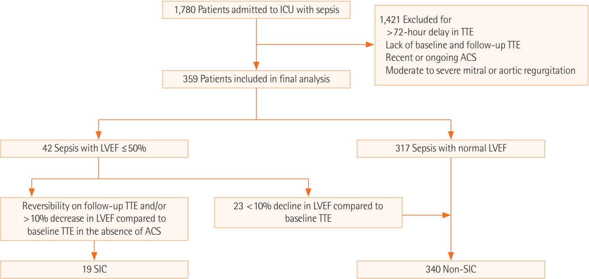
Patient flowchart. ICU: intensive care unit; TTE: transthoracic echocardiogram; ACS: acute coronary syndrome; LVEF: left ventricular ejection fraction; SIC: sepsis-induced cardiomyopathy.
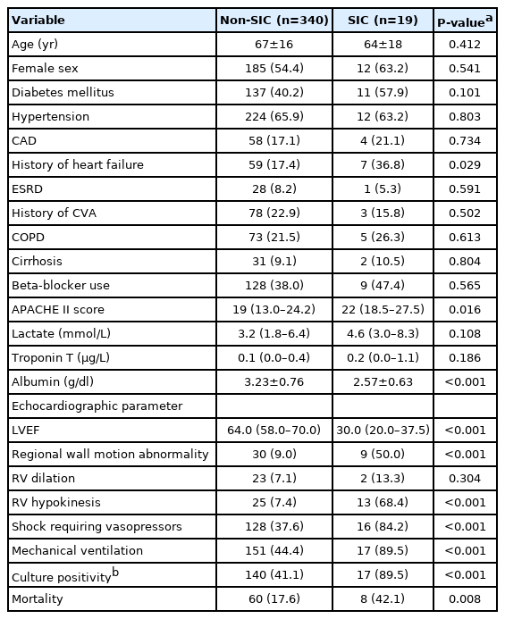
Baseline patient characteristics
Amongst the 359 study participants, the following seven factors were found to be associated with increased all-cause in-hospital mortality in univariable unadjusted analysis: SIC (odds ratio [OR], 4.35; 95% confidence interval [CI], 2.19–8.63; P < 0.01), albumin level (OR, 0.41; 95% CI, 0.29–0.57; P < 0.01), APACHE II (OR, 1.77; 95% CI, 1.04–2.27; P < 0.001), culture positivity (OR, 1.81; 95% CI, 1.10–3.01; P = 0.02), lactate (OR, 1.26; 95% CI, 1.18–1.35; P < 0.001), shock requiring vasopressors (OR, 7.03; 95% CI, 4.05–12.60; P < 0.001), and mechanical ventilation (OR, 9.18; 95% CI, 4.98–18.15; P < 0.001) (Table 2). In the multivariable regression model analysis, after adjusting for albumin level, APACHE II score, culture positivity, lactate, shock requiring vasopressors, and mechanical ventilation, SIC was associated with increased all-cause in-hospital mortality (OR, 4.46; 95% CI, 1.15–18.69; P = 0.03) (Table 2).

Predictors of mortality in patients admitted to the medical intensive care unit with sepsis
Albumin level and culture positivity were independently associated with the development of SIC in the multivariable regression model (Table 3). Each one-unit increase in the albumin level (g/dl) was associated with a lower likelihood of developing SIC (OR, 0.47; CI, 0.23–0.93; P = 0.03). Meanwhile, culture positivity was associated with a greater likelihood of developing SIC (OR, 8.47; CI, 2.24–55.61; P = 0.006). In our cohort, age, history of DM, and cardiac biomarkers were not associated with a greater likelihood of SIC.
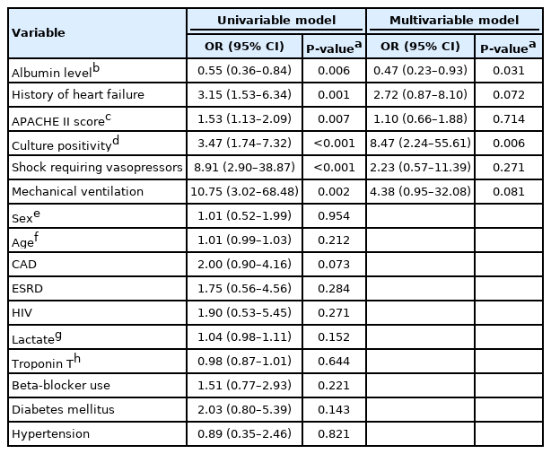
Predictors of SIC in patients admitted to the medical intensive care unit with sepsis
DISCUSSION
Our study evaluated the predictors of SIC and the association of SIC with mortality in sepsis. Our study demonstrates as association between SIC and increased mortality in patients with sepsis, and this association was maintained after adjusting for sepsis severity indices. We also found that low albumin and culture positivity were independently associated with development of SIC. Additionally, we noted biventricular dysfunction was more common than isolated left ventricular (LV) dysfunction in SIC. Lastly, the incidence of SIC was 5.2% among our cohort of patients admitted to the ICU with sepsis.
Prior to 2010, studies on SIC showed that, during sepsis, patients who developed reduced EF were more likely to survive relative to those who continued to have normal or hyperdynamic LV systolic function [3,9,14-16]. This contrasts with our finding of an association between SIC and greater mortality among patients with sepsis. Of course, some possible explanations may account for these differences. First, unlike in our study, the aforementioned previous studies did not adjust for sepsis severity scores in the final analysis of mortality. Second, the first-line vasopressor used in our study was norepinephrine, based on the current sepsis guidelines, whereas earlier studies employed dopamine or epinephrine [3,9,14], which are potentially harmful in a hyperdynamic state, given their higher beta-1 agonist activity.
Biventricular dysfunction was more common than isolated LV dysfunction in patients with SIC. RV hypokinesis, a marker of RV dysfunction, was present in 68% of our SIC patients. This is likely an underestimation of the rate of RV dysfunction. Achieving an accurate estimation of RV dysfunction is challenging as reliable and clinically useful markers for RV function are still under active investigation [17]. Other investigators have previously demonstrated the potential for biventricular dysfunction in SIC, and RV dysfunction rates of up to 52% have been reported in the setting of septic shock [18].
The incidence of SIC was 5.2% in our study evaluating patients with sepsis. This is comparable to rates in other studies that did not limit the study population to only those with shock [6,19]. However, an incidence of up to 60% has been reported by one study focusing on patients with septic shock using daily transesophageal echocardiogram (TEE) [14]. Apart from the aforementioned disparity in patient populations, differences in technique (TEE vs. TTE) and frequency of imaging (daily TEE vs. one-time TTE) may account for the variation in incidence. Our study, along with prior investigations, showed that cardiomyopathy is a common phenomenon in sepsis and that its likelihood increases in patients who progress to shock.
We found that both low albumin level and culture positivity were independently associated with the development of SIC. Albumin, a negative acute-phase reactant, decreases with worsening sepsis and is an indicator of the severity of sepsis [20]. Culture positivity, while it does not directly predict the severity of sepsis, could be representative of the burden of infection [21]. Other factors, such as APACHE II score, mechanical ventilation, and shock requiring vasopressors [22]—which are all indicators of sepsis severity—were found to be significant predictors of SIC in the univariable analysis; however, in the multivariable analysis, the P-values trended towards statistical significance but ultimately did not reach the cutoff value (P<0.05). We believe that the lack of significance was due to the small sample size and inadequate power of this study. We hypothesize that, in a larger study, APACHE II score, mechanical ventilation, shock requiring vasopressors, and a history of heart failure would be independently associated with the development of SIC. Some previously reported factors, such as age and DM, were not identified as predictors of SIC in our cohort. Elevated troponin was also not associated with the development of SIC, consistent with findings from prior research [12]. These results suggest that the severity of sepsis is a stronger predictor of SIC than cardiac risk factors. Thus, factors indicating the severity of sepsis (e.g., low albumin level, APACHE II score) would be more strongly associated with the development of SIC than those indicating cardiac risk (e.g., DM, cardiac biomarkers).
Although our study has a number of novel findings, they need to be interpreted in the context of the following limitations. First, we may have underestimated LV systolic dysfunction by using LVEF as a marker. Using LV end-systolic elastance (LVESE) or global longitudinal strain values may offer a more accurate estimate of LV systolic dysfunction, as these measures do not vary with preload or afterload, both of which are dynamic in the setting of sepsis. LVESE was not assessed in our study and is not routinely assessed clinically. Second, as noted earlier in the discussion, RV dysfunction may also have been underestimated since we used RV hypokinesis as the clinical surrogate. Third, SIC is rapidly reversible over the course of days. Since we only included patients who underwent TTE within 72 hours of diagnosis of sepsis in our study, the incidence of SIC may actually be higher or lower depending on the timing of imaging. Fourth, we employed a multivariable Cox proportional hazards model for analysis and, importantly, two inherent limitations of this method of statistical analysis exist, including that the effect of unknown confounders cannot be accounted for in the study outcome and, due to the small sample size in the SIC group, the possibility of overfitting cannot be excluded.
SIC is a complex reversible cardiomyopathy seen in patients with sepsis and septic shock. Data regarding mortality in SIC have been conflicting. Our study demonstrated that SIC was an independent predictor of mortality in septic patients. Biventricular dysfunction was more common than LV dysfunction alone in patients with SIC. We found that culture positivity and low albumin were independent predictors for the development of SIC, while DM, a history of heart failure, and cardiac biomarkers were not.
KEY MESSAGES
▪ Sepsis-induced cardiomyopathy (SIC) is associated with a higher mortality rate in patients with sepsis, even following adjustment for sepsis severity scores in the final analysis of mortality.
▪ Factors indicating the severity of sepsis (such as low albumin level) rather than those indicating cardiac risk (e.g., diabetes mellitus, cardiac biomarkers) were associated with the development of SIC.
▪ Biventricular dysfunction was more common than isolated left ventricular dysfunction in SIC.
Notes
CONFLICT OF INTEREST No potential conflict of interest relevant to this article was reported.
AUTHOR CONTRIBUTIONS
Conceptualization: BKJH, RTF, PG. Methodology: BKJH, AK (Adarsh Katamreddy), JSG, JYY, RTF, PG. Formal analysis: BKJH, ASN, AK (Adarsh Katamreddy), JYY, PG. Data curation: BKJH, ASN, OLO, AK (Ankit Kushwaha), AK (Ankita Krishnan), MN. Project administration: BKJH, LP, JM, ST, SM, AG, HG, MS, RTF, PG. Visualization: BKJH, PG. Writing - Original draft: ASN, AK (Adarsh Katamreddy), JSG, JYY, MS, PG. Writing - Review and editing: BKJH, ASN, AK (Adarsh Katamreddy), OLO, AK (Ankit Kushwaha), AK (Ankita Krishnan), MN, LP, JM, ST, SM, AG, HG, MS, RTF, PG. Writing–review & editing: all authors.