Contrast media mimicking subarachnoid hemorrhage after intrathecal injection in a patient with Creutzfeldt-Jakob disease
Article information
Acute Crit Care. 2022;37(4):693-693
Publication date (electronic) : 2022 November 30
doi :
https://doi.org/10.4266/acc.2022.00339.e1
Received 2022 September 30; Revised 2022 September 30; Accepted 2022 September 30.
Acute and Critical Care 2022;37:474-476
https://doi.org/10.4266/acc.2022.00339
In the article entitled “Contrast media mimicking subarachnoid hemorrhage after intrathecal injection in a patient with Creutzfeldt-Jakob disease”, the figure legend was incorrectly presented. The correct figure is as follows.
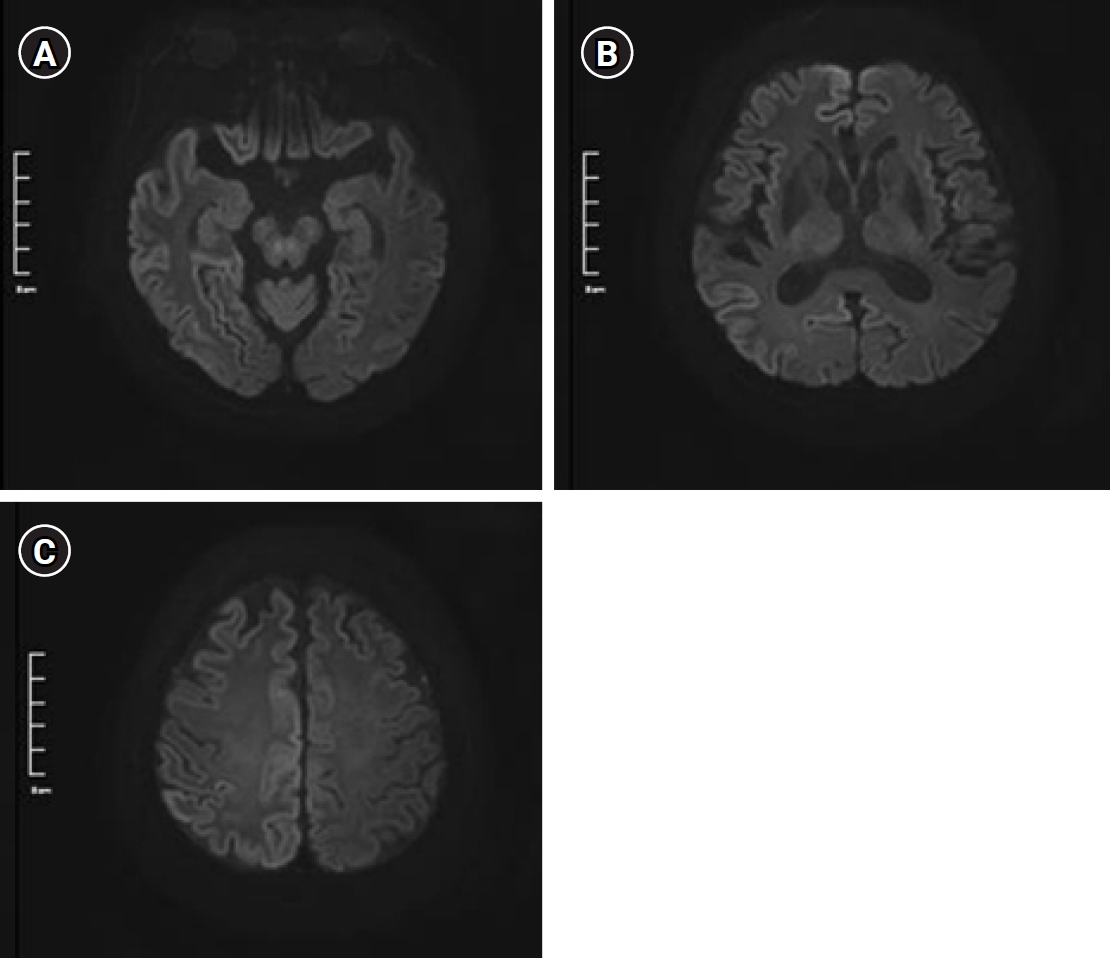
Brain diffusion-weighted imaging at b1000 at the levels of (A) midbrain, (B) thalamus, and (C) frontal and parietal lobes. Multifocal diffusion restriction can be seen along the bilateral cerebral cortex, especially prominent on the right parietal area.
Article information Continued
Copyright © 2022 The Korean Society of Critical Care Medicine