Skip Navigation
Skip to contents

ACC : Acute and Critical Care

OPEN ACCESS
SEARCH
Search

Articles

Page Path
HOME > Acute Crit Care > Volume 36(3); 2021 > Article
Original Article
Pulmonary
Effects of high-flow nasal cannula in patients with mild to moderate hypercapnia: a prospective observational study
Kyung Hun Nam1,*orcid, Hyung Koo Kang2orcid, Sung-Soon Lee2orcid, So-Hee Park2orcid, Sung Wook Kang3orcid, Jea Jun Hwang3orcid, So Young Park4orcid, Won Young Kim5orcid, Hee Jung Suh6orcid, Eun Young Kim6orcid, Ga Jin Seo6orcid, Younsuck Koh7orcid, Sang-Bum Hong7orcid, Jin Won Huh7orcid, Chae-Man Lim7orcid
Acute and Critical Care 2021;36(3):249-255.
DOI: https://doi.org/10.4266/acc.2020.01102
Published online: July 26, 2021

1Department of Medicine, Ajou University Intermediate Care Hospital, Suwon, Korea

2Division of Pulmonary and Critical Care Medicine, Department of Internal Medicine, Inje University Ilsan Paik Hospital, Inje University College of Medicine, Goyang, Korea

3Department of Pulmonary and Critical Care Medicine, Kyung Hee University Hospital at Gangdong, Seoul, Korea

4Department of Pulmonology, Ewha Womans University Seoul Hospital, Seoul, Korea

5Department of Emergency Medicine, Asan Medical Center, University of Ulsan College of Medicine, Seoul, Korea

6Intensive Care Nursing Team, Asan Medical Center, Seoul, Korea

7Department of Pulmonary and Critical Care Medicine, Asan Medical Center, University of Ulsan College of Medicine, Seoul, Korea

Corresponding author Chae-Man Lim Department of Pulmonary and Critical Care Medicine, Asan Medical Center, University of Ulsan College of Medicine, 88 Olympic-ro 43-gil, Songpa-gu, Seoul 05505, Korea Tel: +82-2-3010-4700 Fax: +82-2-2045-4039 E-mail: cmlim@amc.seoul.kr
*Current affiliation: Department of Critical Care Medicine, Seongnam Citizens Medical Center, Seongnam, Korea
• Received: December 10, 2020   • Revised: April 19, 2021   • Accepted: April 19, 2021

Copyright © 2021 The Korean Society of Critical Care Medicine

This is an Open Access article distributed under the terms of the Creative Commons Attribution Non-Commercial License (http://creativecommons.org/licenses/by-nc/4.0/) which permits unrestricted non-commercial use, distribution, and reproduction in any medium, provided the original work is properly cited.

  • 7,261 Views
  • 277 Download
  • 5 Web of Science
  • 6 Crossref
  • 4 Scopus
prev next
  • Background
    Evidence for using high-flow nasal cannula (HFNC) in hypercapnia is still limited. Most of the clinical studies had been conducted retrospectively, and there had been conflicting reports for the effects of HFNC on hypercapnia correction in prospective studies. Therefore, more evidence is needed to understand the effect of the HFNC in hypercapnia.
  • Methods
    We conducted a multicenter prospective observational study after applying HFNC to 45 hospitalized subjects who had moderate hypercapnia (arterial partial pressure of carbon dioxide [PaCO2], 43–70 mm Hg) without severe respiratory acidosis (pH <7.30). The primary outcome was a change in PaCO2 level in the first 24 hours of HFNC use. The secondary outcomes were changes in other parameters of arterial blood gas analysis, changes in respiration rates, and clinical outcomes.
  • Results
    There was a significant decrease in PaCO2 in the first hour of HFNC application (-3.80 mm Hg; 95% confidence interval, -6.35 to -1.24; P<0.001). Reduction of PaCO2 was more prominent in subjects who did not have underlying obstructive lung disease. There was a correction in pH, but no significant changes in respiratory rate, bicarbonate, and arterial partial pressure of oxygen/fraction of inspired oxygen ratio. Mechanical ventilation was not required for 93.3% (42/45) of our study population.
  • Conclusions
    We suggest that HFNC could be a safe alternative for oxygen delivery in hypercapnia patients who do not need immediate mechanical ventilation. With HFNC oxygenation, correction of hypercapnia could be expected, especially in patients who do not have obstructive lung diseases.
High-flow nasal cannula (HFNC) is a relatively new device for oxygenation that has become a popular tool in the treatment of hypoxic respiratory failure [1]. Recently, there has been a growing interest in its use in hypercapnia. Physiological studies have shown that HFNC reduces rebreathing of carbon dioxide in the upper and lower anatomical dead space [2,3]. HFNC users generate less carbon dioxide by reduced work of breathing [4]. Some studies have also suggested that HFNC may even improve alveolar ventilation [5]. However, the effectiveness of HFNC in hypercapnic respiratory failure has not been fully demonstrated as relevant studies have been mostly retrospective with some contradictory results [6-8].
To clearly understand the effects of HFNC in hypercapnic respiratory failure, we conducted a prospective observational study with frequent arterial blood gas analysis (ABGA) in the first 24 hours after starting HFNC. For safety reasons, we recruited patients with mild to moderate hypercapnia without severe acidosis. The aim of this study was to examine the competence of HFNC in hypercapnia correction and to investigate the clinical use of HFNC in patients with hypercapnia.
A multicenter prospective observational study was conducted in four tertiary care hospitals in South Korea from November 2017 to October 2018. Inclusion criteria for the subjects were patients with mild to moderate hypercapnia (43 ≤ arterial partial pressure of carbon dioxide [PaCO2] ≤ 70 mm Hg) in ABGA. Recruitment was conducted from the emergency room, general ward, and intensive care unit. Exclusion criteria were: under 18 years of age; respiratory rate more than 35 breaths per minute; severe acidosis (pH < 7.30); using accessory muscle of respiration; any other signs of progressive respiratory failure requiring imminent intubation and mechanical ventilation; unstable hemodynamics; post-cardiopulmonary resuscitation status or on extracorporeal membrane oxygenation; consciousness disorder that would require intubation (e.g., not responsive, agitated, uncooperative, high risk for aspiration); facial surgery, trauma or deformity and airway obstruction that precluded application of nasal cannula; and those receiving end-of-life care.
HFNC was initiated with a pulse oximeter oxygen saturation target of 92%. The initial flow was set at 35 L/min and adjusted to suit the best comfort of the subject. ABGA was checked at 1, 3, 6, and 24 hours after starting HFNC. When the 24-hour data collecting period ended, the attending physician decided on the continuation and duration of the HFNC. Medications such as bronchodilators and antibiotics were administered to treat the underlying medical condition and precipitating factors.
HFNC was discontinued if the subject met any of the following criteria: symptoms and signs of worsening respiratory distress, such as retractions or sweating; increased respiratory rate to more than 35 breaths per minute; elevation of PaCO2 greater than 10 mm Hg from the previous ABGA results; pH less than 7.2 in any of the ABGA results; pH decrease more than 0.2 from previous ABGA results; or deteriorating mental status.
Study Outcomes
The primary outcome was change in ABGA parameters during the first 24 hours of HFNC application. We also analyzed change in PaCO2 based on underlying lung conditions. We hypothesized that the reduction in PaCO2 would be greater in patients with an obstructive pulmonary pathology. The secondary outcome was the clinical course of the subjects during hospitalization, which included discontinuation of HFNC according to the criteria, progression to invasive or noninvasive mechanical ventilation, and death.
Statistical Analysis
One-way repeated measure analysis of variance with a post hoc test by Bonferroni correction was used to evaluate the changes in ABGA parameters. Descriptive statistics were used for the clinical courses of subjects. The level of significance was set at 0.05. IBM SPSS ver. 25 (IBM Corp., Armonk, NY, USA) was used for all analyses.
Ethics Statement
The Institutional Review Boards of each participating hospital approved the study and written informed consent was obtained from all study participants prior to initial observation. This study is registered in the Korean Clinical Research Information Service (CRIS, https://cris.nih.go.kr; registration No. KCT0003448).
Study Population
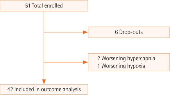
A total of 51 subjects were recruited for the study. Six subjects dropped out of the study due to discomfort associated with HFNC (Figure 1). The general characteristics of the 45 study subjects are reported (Table 1). The most common acute lung condition was pneumonia followed by pulmonary edema and chronic obstructive pulmonary disease (COPD) exacerbation. In total, 87% (39/45) of the patients were receiving conventional oxygen therapy at the time of enrolment. For the initial HFNC settings, the median fraction of inspired oxygen (FiO2) provided was 32% with a median flow of 35 L/min.
Primary Outcome
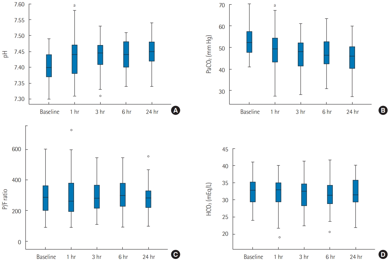
Changes in ABGA parameters are illustrated (Figure 2). There were significant pH and PaCO2 changes after HFNC application (Table 2). For PaCO2, a significant decrease was observed between the baseline and 1-hour measurement (–3.80 mm Hg; 95% confidence interval [CI], –6.35 to –1.24; P < 0.001). Among other checkpoints, there were no significant differences. Similarly, for pH, a significant increase was observed between the baseline and 1-hour measurement (0.030; 95% CI, 0.050–0.010; p < 0.001). In other parameters such as arterial bicarbonate concentration (HCO3) and arterial partial pressure of oxygen/fraction of inspired oxygen ratio, no significant differences were observed between checkpoints.

PaCO2 changes in the obstructive and non-obstructive groups

We analyzed changes in PaCO2 after grouping the subjects based on underlying lung diseases. The “obstructive group” included subjects who had previously been diagnosed with obstructive lung disease: COPD, chronic bronchitis, emphysema, bronchiectasis and asthma. The “non-obstructive group” included subjects who had no documented obstructive lung disease. There were 18 subjects in the obstructive group and 24 in the non-obstructive group. Between the two groups, there was no statistical difference in age, sex, BMI, baseline ABGA results, and the initial HFNC setting.
PaCO2 changes in separate groups are illustrated (Figure 3). In the obstructive group, mean PaCO2 decreased from 54.2 to 51.8, 49.7, 49.6, and 48.3 mm Hg. These changes were not statistically significant (P=0.075). However, a significant decrease of PaCO2 was also found in the non-obstructive group. As mean PaCO2 decreased from 51.7 to 46.9, 46.0, 44.8, and 44.2 mm Hg, there was a significant decrease between baseline and 1-hour measurement (–4.83 mm Hg; 95% CI, –8.4 to –1.3; P < 0.001).
Secondary Outcome
Among the 45 study subjects, three subjects (6.7%) met the criteria for HFNC discontinuation. Two patients had an increase in PaCO2 of more than 10 mm Hg, and one patient showed symptoms and signs of worsening respiratory distress. Noninvasive ventilation was applied to the two patients with worsening hypercapnia. Invasive mechanical ventilation was applied to the patient with worsening respiratory distress. For the remainder of the 42 subjects, HFNC was continued for an average of 47 hours (median, 33 hours; interquartile range, 24–70 hours). No deaths were observed among the study population.
In this prospective observational study, we report our findings on changes in ABGA parameters after applying HFNC to patients with mild to moderate hypercapnia. A significant decrease in PaCO2 and pH correction were observed in the first hour of HFNC application. The effect of PaCO2 reduction was more prominent in patients who did not have underlying obstructive lung disease. In terms of clinical outcomes of the 45 study subjects, two subjects required noninvasive mechanical ventilation for worsening hypercapnia, and one subject required invasive mechanical ventilation after intubation for worsening respiratory distress. There were no deaths or serious adverse outcomes in the study population.
With its unique physiological benefits, HFNC has become a popular device for oxygen delivery. HFNC provides a constant FiO2, improves mucociliary clearance, reduces the effort required to breathe, and decreases anatomical dead space [1]. With acute hypoxic respiratory failure, multiple prospective randomized trials have shown the clinical advantages of HFNC over conventional oxygen delivery systems [9,10]. Therefore, HFNC is widely being used in various conditions with hypoxia.
However, the effects of HFNC on hypercapnic respiratory failure remain unclear; therefore, more evidence is needed to support its use in patients with hypercapnia. Although most clinical studies have reported a reduction in PaCO2 after applying HFN, there have been some studies with conflicting results. In a study by Sztrymf et al. [8], involving intensive care unit patients with acute respiratory failure, a significant increase in PaCO2 was observed. In a study by Nilius et al. [11], the use of HFNC resulted in different individual responses in respiratory rates and PaCO2 levels.
Because hypercapnia has complex pathophysiological mechanisms and multiple etiologies, there is wide heterogeneity in patients [12]. This could explain the mixed results in clinical studies and becomes a challenge to establishing clear indications for the use of HFNC in hypercapnic respiratory failure. However, even with various causes, there are commonalities in these patients. Whatever the diagnosis, mechanical ventilation (invasive and noninvasive) is a standard treatment in severe cases of hypercapnia [13]. Therefore, the potential CO2 removal capacity and physiologic benefits make HFNC an attractive alternative to conventional oxygen therapy in patients with hypercapnia.
With our study, an overall decrease in PaCO2 with a correction of pH was observed after applying HFNC in patients with mild to moderate hypercapnia. Additionally, 93% of the study population was successfully treated without progressing to mechanical ventilation. In other studies investigating clinical implications of HFNC on hypercapnic respiratory failure, intubation rate ranged from 0% to 25% [14,15]. A recent metaanalysis suggested that HFNC was not inferior to noninvasive ventilation with respect to intubation rate and mortality [16]. Considering all current evidence, HFNC may be a feasible alternative option for oxygen supplementation in patients with mild to moderate hypercapnia.
However, our study found a different response to HFNC depending on underlying lung disease. We analyzed changes in PaCO2 after grouping the patients according to the presence or absence of an obstructive lung pathology. Contrary to our anticipation, the effects of HFNC were more pronounced in patients who did not have underlying obstructive lung disease. This finding suggests that the decrease in PaCO2 during HFNC was possibly related to the decreased effort required to breathe rather than reduction in dead space. Further studies are needed to better understand the effects of HFNC on hypercapnia based on underlying lung pathology.
We recognize the limitations of this study. We acknowledge that the findings in this study cannot be fully accepted without a control group comparison. Also, because of the nature of this study design, selection bias is possible. Last, the small number of subjects also limits the ability to generalize the study’s findings.
In conclusion, we prospectively investigated changes in ABGA parameters and clinical implications of HFNC in patients with mild to moderate hypercapnia. After applying HFNC, an overall decrease in PaCO2 with pH correction was seen. Most of our subjects were treated successfully without progressing to mechanical ventilation. Therefore, we suggest that HFNC could be a feasible alternative option for oxygen delivery in patients with mild to moderate hypercapnia. However, further controlled studies with different etiologies of hypercapnia are needed to establish the clear role of HFNC in patients with hypercapnic respiratory failure.
■ When high-flow nasal cannula was applied in moderate hypercapnia patients without severe respiratory acidosis, reduction of carbon dioxide and correction of pH was seen.
■ Progression to mechanical ventilation was comparable to previous studies.
■ We suggest that high-flow nasal cannula could be a safe alternative for oxygen delivery in hypercapnia patients who do not need immediate mechanical ventilation.

CONFLICT OF INTEREST No potential conflict of interest relevant to this article was reported.

AUTHOR CONTRIBUTIONS

Conceptualization: KHN, WYK, CML. Data curation: KHN, HJS, EYK, GJS. Formal analysis: KHN. Funding acquisition: CML. Methodology: all authors. Project administration: CML, KHN. Investigation: all authors. Visualization: KHN. Writing–original draft: KHN. Writing–review & editing: CML.

Funding was provided by the Korea Health Industry Development Institute (https://www.khidi.or.kr; grant No. HI17C0263). High-flow nasal cannula device was sponsored by MEK-ICS, South Korea (http://mek-ics.com). Neither had a role in the study.
Figure 1.
Flow of study participants.
acc-2020-01102f1.jpg
Figure 2.
Change in arterial blood gas analysis parameters at baseline, 1, 3, 6, and 24 hours after applying high-flow nasal cannula. (A) pH. (B) Arterial partial pressure of carbon dioxide (PaCO2). (C) Arterial partial pressure of oxygen/fraction of inspired oxygen (PaO2/FiO2) ratio (P/F ratio). (D) Arterial bicarbonate concentration (HCO3). aStatistically significant change from previous measurement.
acc-2020-01102f2.jpg
Figure 3.
Change in arterial partial pressure of carbon dioxide (PaCO2) at baseline, 1, 3, 6, and 24 hours after applying high-flow nasal cannula. (A) Obstructive group. (B) Non-obstructive group. aStatistically significant change from previous measurement.
acc-2020-01102f3.jpg
Table 1.
General characteristics of enrolled subjects
Characteristics Total (n=45)
Demographics
 Age (yr) 70 (62–81)
 Male sex 27 (60.0)
 BMI (kg/m2) 21.3 (18.0–25.2)
Acute lung condition
 Pneumonia 23 (48.9)
 Pulmonary edema 15 (33.3)
 COPD exacerbation 12 (26.7)
 Atelectasis 6 (13.3)
 Asthma exacerbation 3 (6.7)
Baseline conventional O2
 Nasal prong 32 (71.1)
 Face mask 7 (15.6)
Initial HFNC setting
 FiO2 (%) 32 (25–37)
 Flow (L/min) 35 (30–37.5)

Values are presented as median (interquartile range) or number (%).

BMI: body mass index; COPD: chronic obstructive pulmonary disease; HFNC: high-flow nasal cannula oxygen therapy; FiO2: fraction of inspired oxygen.

Table 2.
Changes in parameters of arterial blood gas analysis after high-flow nasal cannula application
Variable Baseline 1 hr 3 hr 6 hr 24 hr P-value
pH 7.402±0.05 7.431±0.06 7.436±0.05 7.435±0.05 7.444±0.05 <0.001
PaCO2 (mm Hg) 52.8±6.8 49.0±9.0 47.6±7.0 46.9±7.0 45.9±8.3 <0.001
P/F ratio 99±93 294±134 288±108 296±105 283±96 0.826
HCO3 (mEq/L) 31.8±5.3 32.2±5.0 32.0±4.4 31.6±4.5 31.4±4.5 0.518

Values are presented as mean±standard deviation.

hr: hours after high-flow nasal cannula application; PaCO2: arterial partial pressure of carbon dioxide; P/F ratio: arterial partial pressure of oxygen/fraction of inspired oxygen (PaO2/FiO2) ratio; HCO3: arterial bicarbonate concentration.

  • 1. Drake MG. High-flow nasal cannula oxygen in adults: an evidence-based assessment. Ann Am Thorac Soc 2018;15:145-55.ArticlePubMed
  • 2. Möller W, Celik G, Feng S, Bartenstein P, Meyer G, Oliver E, et al. Nasal high flow clears anatomical dead space in upper airway models. J Appl Physiol (1985) 2015;118:1525-32.ArticlePubMedPMC
  • 3. Bräunlich J, Goldner F, Wirtz H. Nasal highflow eliminates CO2 from lower airways. Respir Physiol Neurobiol 2017;242:86-8.ArticlePubMed
  • 4. Biselli PJ, Kirkness JP, Grote L, Fricke K, Schwartz AR, Smith P, et al. Nasal high-flow therapy reduces work of breathing compared with oxygen during sleep in COPD and smoking controls: a prospective observational study. J Appl Physiol (1985) 2017;122:82-8.ArticlePubMed
  • 5. Patel A, Nouraei SA. Transnasal humidified rapid-insufflation ventilatory exchange (THRIVE): a physiological method of increasing apnoea time in patients with difficult airways. Anaesthesia 2015;70:323-9.ArticlePubMed
  • 6. Jeong JH, Kim DH, Kim SC, Kang C, Lee SH, Kang TS, et al. Changes in arterial blood gases after use of high-flow nasal cannula therapy in the ED. Am J Emerg Med 2015;33:1344-9.ArticlePubMed
  • 7. Kim ES, Lee H, Kim SJ, Park J, Lee YJ, Park JS, et al. Effectiveness of high-flow nasal cannula oxygen therapy for acute respiratory failure with hypercapnia. J Thorac Dis 2018;10:882-8.ArticlePubMedPMC
  • 8. Sztrymf B, Messika J, Mayot T, Lenglet H, Dreyfuss D, Ricard JD. Impact of high-flow nasal cannula oxygen therapy on intensive care unit patients with acute respiratory failure: a prospective observational study. J Crit Care 2012;27:324.e9-13.ArticlePubMed
  • 9. Frat JP, Thille AW, Mercat A, Girault C, Ragot S, Perbet S, et al. High-flow oxygen through nasal cannula in acute hypoxemic respiratory failure. N Engl J Med 2015;372:2185-96.ArticlePubMed
  • 10. Rochwerg B, Granton D, Wang DX, Helviz Y, Einav S, Frat JP, et al. High flow nasal cannula compared with conventional oxygen therapy for acute hypoxemic respiratory failure: a systematic review and meta-analysis. Intensive Care Med 2019;45:563-72.ArticlePubMed
  • 11. Nilius G, Franke KJ, Domanski U, Rühle KH, Kirkness JP, Schneider H. Effects of nasal insufflation on arterial gas exchange and breathing pattern in patients with chronic obstructive pulmonary disease and hypercapnic respiratory failure. Adv Exp Med Biol 2013;755:27-34.ArticlePubMed
  • 12. Roussos C, Koutsoukou A. Respiratory failure. Eur Respir J Suppl 2003;47:3s-14s.ArticlePubMed
  • 13. Davidson AC, Banham S, Elliott M, Kennedy D, Gelder C, Glossop A. BTS/ICS guideline for the ventilatory management of acute hypercapnic respiratory failure in adults. Thorax 2016;71 Suppl 2:ii1-35.ArticlePubMed
  • 14. Papachatzakis Y, Nikolaidis PT, Kontogiannis S, Trakada G. High-flow oxygen through nasal cannula vs. non-invasive ventilation in hypercapnic respiratory failure: a randomized clinical trial. Int J Environ Res Public Health 2020;17:5994. ArticlePubMedPMC
  • 15. Lee MK, Choi J, Park B, Kim B, Lee SJ, Kim SH, et al. High flow nasal cannulae oxygen therapy in acute-moderate hypercapnic respiratory failure. Clin Respir J 2018;12:2046-56.ArticlePubMed
  • 16. Huang Y, Lei W, Zhang W, Huang JA. High-flow nasal cannula in hypercapnic respiratory failure: a systematic review and meta-analysis. Can Respir J 2020;2020:7406457. ArticlePubMedPMC

Figure & Data

References

    Citations

    Citations to this article as recorded by  
    • A high-flow nasal cannula versus noninvasive ventilation in acute exacerbations of chronic obstructive pulmonary disease
      Oguzhan Haciosman, Huseyin Ergenc, Adem Az, Yunus Dogan, Ozgur Sogut
      The American Journal of Emergency Medicine.2025; 87: 38.     CrossRef
    • Oxygénothérapie nasale à haut débit et insuffisance respiratoire aiguë hypercapnique
      C. Girault, E. Artaud-Macari, G. Jolly, D. Carpentier, A. Cuvelier, G. Béduneau
      Revue des Maladies Respiratoires.2024; 41(7): 498.     CrossRef
    • Safety and efficacy of high flow nasal canula in patients with mild hypercapnia
      Mohammed A. Ibrahim, Magdy Emara, Mohammed Shehta
      The Egyptian Journal of Bronchology.2023;[Epub]     CrossRef
    • Current status of treatment of acute respiratory failure in Korea
      Yong Jun Choi, Jae Hwa Cho
      Journal of the Korean Medical Association.2022; 65(3): 124.     CrossRef
    • High-flow nasal cannula: Evaluation of the perceptions of various performance aspects among Chinese clinical staff and establishment of a multidimensional clinical evaluation system
      Ruoxuan Wen, Xingshuo Hu, Tengchen Wei, Kaifei Wang, Zhimei Duan, Zhanqi Zhao, Lixin Xie, Fei Xie
      Frontiers in Medicine.2022;[Epub]     CrossRef
    • The Application Progress of HFNC in Respiratory Diseases
      迪 吴
      Advances in Clinical Medicine.2022; 12(11): 10617.     CrossRef

    • PubReader PubReader
    • ePub LinkePub Link
    • Cite
      CITE
      export Copy
      Close
      Download Citation
      Download a citation file in RIS format that can be imported by all major citation management software, including EndNote, ProCite, RefWorks, and Reference Manager.

      Format:
      • RIS — For EndNote, ProCite, RefWorks, and most other reference management software
      • BibTeX — For JabRef, BibDesk, and other BibTeX-specific software
      Include:
      • Citation for the content below
      Effects of high-flow nasal cannula in patients with mild to moderate hypercapnia: a prospective observational study
      Acute Crit Care. 2021;36(3):249-255.   Published online July 26, 2021
      Close
    • XML DownloadXML Download
    Figure
    • 0
    • 1
    • 2
    Related articles
    Effects of high-flow nasal cannula in patients with mild to moderate hypercapnia: a prospective observational study
    Image Image Image
    Figure 1. Flow of study participants.
    Figure 2. Change in arterial blood gas analysis parameters at baseline, 1, 3, 6, and 24 hours after applying high-flow nasal cannula. (A) pH. (B) Arterial partial pressure of carbon dioxide (PaCO2). (C) Arterial partial pressure of oxygen/fraction of inspired oxygen (PaO2/FiO2) ratio (P/F ratio). (D) Arterial bicarbonate concentration (HCO3). aStatistically significant change from previous measurement.
    Figure 3. Change in arterial partial pressure of carbon dioxide (PaCO2) at baseline, 1, 3, 6, and 24 hours after applying high-flow nasal cannula. (A) Obstructive group. (B) Non-obstructive group. aStatistically significant change from previous measurement.
    Effects of high-flow nasal cannula in patients with mild to moderate hypercapnia: a prospective observational study
    Characteristics Total (n=45)
    Demographics
     Age (yr) 70 (62–81)
     Male sex 27 (60.0)
     BMI (kg/m2) 21.3 (18.0–25.2)
    Acute lung condition
     Pneumonia 23 (48.9)
     Pulmonary edema 15 (33.3)
     COPD exacerbation 12 (26.7)
     Atelectasis 6 (13.3)
     Asthma exacerbation 3 (6.7)
    Baseline conventional O2
     Nasal prong 32 (71.1)
     Face mask 7 (15.6)
    Initial HFNC setting
     FiO2 (%) 32 (25–37)
     Flow (L/min) 35 (30–37.5)
    Variable Baseline 1 hr 3 hr 6 hr 24 hr P-value
    pH 7.402±0.05 7.431±0.06 7.436±0.05 7.435±0.05 7.444±0.05 <0.001
    PaCO2 (mm Hg) 52.8±6.8 49.0±9.0 47.6±7.0 46.9±7.0 45.9±8.3 <0.001
    P/F ratio 99±93 294±134 288±108 296±105 283±96 0.826
    HCO3 (mEq/L) 31.8±5.3 32.2±5.0 32.0±4.4 31.6±4.5 31.4±4.5 0.518
    Table 1. General characteristics of enrolled subjects

    Values are presented as median (interquartile range) or number (%).

    BMI: body mass index; COPD: chronic obstructive pulmonary disease; HFNC: high-flow nasal cannula oxygen therapy; FiO2: fraction of inspired oxygen.

    Table 2. Changes in parameters of arterial blood gas analysis after high-flow nasal cannula application

    Values are presented as mean±standard deviation.

    hr: hours after high-flow nasal cannula application; PaCO2: arterial partial pressure of carbon dioxide; P/F ratio: arterial partial pressure of oxygen/fraction of inspired oxygen (PaO2/FiO2) ratio; HCO3: arterial bicarbonate concentration.


    ACC : Acute and Critical Care
    TOP