Skip Navigation
Skip to contents

ACC : Acute and Critical Care

OPEN ACCESS
SEARCH
Search

Articles

Page Path
HOME > Acute Crit Care > Volume 39(2); 2024 > Article
Review Article
Infection
Microbial infections in burn patients
Souvik Roy1orcid, Preeti Mukherjee1orcid, Sutrisha Kundu1orcid, Debashrita Majumder1orcid, Vivek Raychaudhuri1orcid, Lopamudra Choudhury2orcid
Acute and Critical Care 2024;39(2):214-225.
DOI: https://doi.org/10.4266/acc.2023.01571
Published online: May 24, 2024

1Postgraduate and Research Department of Biotechnology, St. Xavier’s College (Autonomous), Kolkata, India

2Department of Microbiology, Sarsuna College (under Calcutta University), Kolkata, India

Corresponding Author: Souvik Roy Postgraduate and Research Department of Biotechnology, St. Xavier’s College (Autonomous), 30, Mother Teresa Sarani, Kolkata 700016, West Bengal, India Email: souvikroybiotech@sxccal.edu
• Received: December 8, 2023   • Revised: March 16, 2024   • Accepted: March 27, 2024

© 2024 The Korean Society of Critical Care Medicine

This is an Open Access article distributed under the terms of the Creative Commons Attribution Non-Commercial License (http://creativecommons.org/licenses/by-nc/4.0/) which permits unrestricted non-commercial use, distribution, and reproduction in any medium, provided the original work is properly cited.

  • 893 Views
  • 94 Download
  • Polymicrobial infections are the leading causes of complications incurred from injuries that burn patients develop. Such patients admitted to the hospital have a high risk of developing hospital-acquired infections, with longer patient stays leading to increased chances of acquiring such drug-resistant infections. Acinetobacter baumannii, Klebsiella pneumoniae, Pseudomonas aeruginosa, and Proteus mirabilis are the most common multidrug-resistant (MDR) Gram-negative bacteria identified in burn wound infections (BWIs). BWIs caused by viruses, like herpes simplex and varicella zoster, and fungi-like Candida species appear to occur occasionally. However, the preponderance of infection by opportunistic pathogens is very high in burn patients. Variations in the causative agents of BWIs are due to differences in geographic location and infection control measures. Overall, burn injuries are characterized by elevated serum cytokine levels, systemic immune response, and immunosuppression. Hence, early detection and treatment can accelerate the wound-healing process and reduce the risk of further infections at the site of injury. A multidisciplinary collaboration between burn surgeons and infectious disease specialists is also needed to properly monitor antibiotic resistance in BWI pathogens, help check the super-spread of MDR pathogens, and improve treatment outcomes as a result.
The human skin is the largest anatomical organ involved in various physiological functions like thermoregulation, maintaining homeostasis, proprioception, and protection from external agents [1]. The skin is man’s physical barrier to resist pathogen attack. Conditions that lead to loss of skin integrity therefore have numerous serious consequences [1].
Burn injury is a major global public health crisis. It disrupts the epidermal barrier, leading to down-regulation of both local and systemic immune responses [1]. As a result, burn wounds become an ideal breeding ground for microbes [1,2]. The burn wound serves as an ideal microenvironment predominated with biological fluids called burn wound exudates (BWEs), which collectively create a perfect niche for the growth of pathogens [3].
First-degree (superficial) burns damage only the epidermal layer, so they heal rather quickly without scarring [2]. Second-degree (partial-thickness) burns involve the deeper layers of the epidermis and dermis and heal slowly [2]. Third-degree (full-thickness) burns fully destroy the epidermal and dermal layers of the skin and can also cause significant damage to the underlying tissues and bones as well [2].
Burn is a very common and devastating form of trauma. It has been ranked seventh among all traumatic injuries by the World Health Organization, with a crude mortality rate of 5% [4]. Across the world, around 2.65 lakh deaths occur every year due to burn injuries. Such cases are more prevalent in developing and under-developed countries, and in these cases, patient mortality potential soars up to 100% with burns covering more than 40% of the total body surface area [3,5]. Around 80% of burns occur at home [6]. Domestic burn injuries are more common among children and adolescents [6,7].
Asia records the highest number of intentional burn injuries in the world, with Southeast Asia topping the list, followed by Africa [8]. Among the Asian countries, India records the highest number of cases of intentional self-harm by burning, followed by Pakistan, Bhutan, and Bangladesh. Africa records the highest mortality rate from burn injuries, 23.5% per year [8]. In India, 65% of the burn victims are young women, due to self-immolation or domestic violence [9]. On the other hand, in Africa, children are the predominant victims of burn injuries. Prevention of burn injuries in Asia and Africa is hampered due to the high population density, lack of education, low income rate, and poor surveillance systems [9].
Reported cases of burn injuries are significantly lower in continents like North America, South America, Australia, and Europe [8]. The victims of intentional self-harm by burning in Europe are more prevalent among men in the age group of 40–50 years [9]. Australia records the highest number of admissions of burn patients in hospitals each year, followed by Asia. These developed continents are in a much better situation concerning burn injuries compared to the under-developed and developing countries. Polymicrobial infections are responsible for 75% of all deaths from burns [2]. The risk factors influencing microbial infections at the burn site include the size and surface area of the burn, age, immune status, the degree of burn, and comorbidities [2].
Burns occur at temperatures above 44 °C [10]. Trans-epidermal necrosis happens in just a second at 70 °C, while it happens in 45 minutes at 47 °C [10]. Fire flaming and scalding represent 23.8% and 66.2% of burn injury cases, respectively [11]. The remaining 10% of burns have other causes [11]. Scalding causes first or second-degree burns, while flame causes second or third-degree burns [10].
Burns are grouped as thermal, chemical, frostbite, electrical, radiation, or sunburn [10]. Around 3%–6% of all burn cases constitute chemical burns, accounting for 14%–30% of mortalities [10]. Chemical burns develop due to contact with coal tar, strong acids, alkaline solutions, or phosphorus due to bomb explosions [10]. Cold burn or frostbite occurs as the skin starts freezing from –10 °C, with irreversible changes occurring below –22 °C [10].
Following burns, microorganisms colonize and grow quickly at the site of injury due to the loss of the skin barrier. The skin barrier otherwise serves as the first line of immune defense for any individual [12-14]. Any breach in the skin allows for easy entry and access of the infecting microbe to the inner tissues of the body, thus complicating the etiology [12-14]. Hence, it has been observed that microbial infections, especially those caused by multidrug-resistant (MDR)-bacteria, including Pseudomonas and Acinetobacter, are the main cause of increased morbidity and mortality in burn patients [12-14].
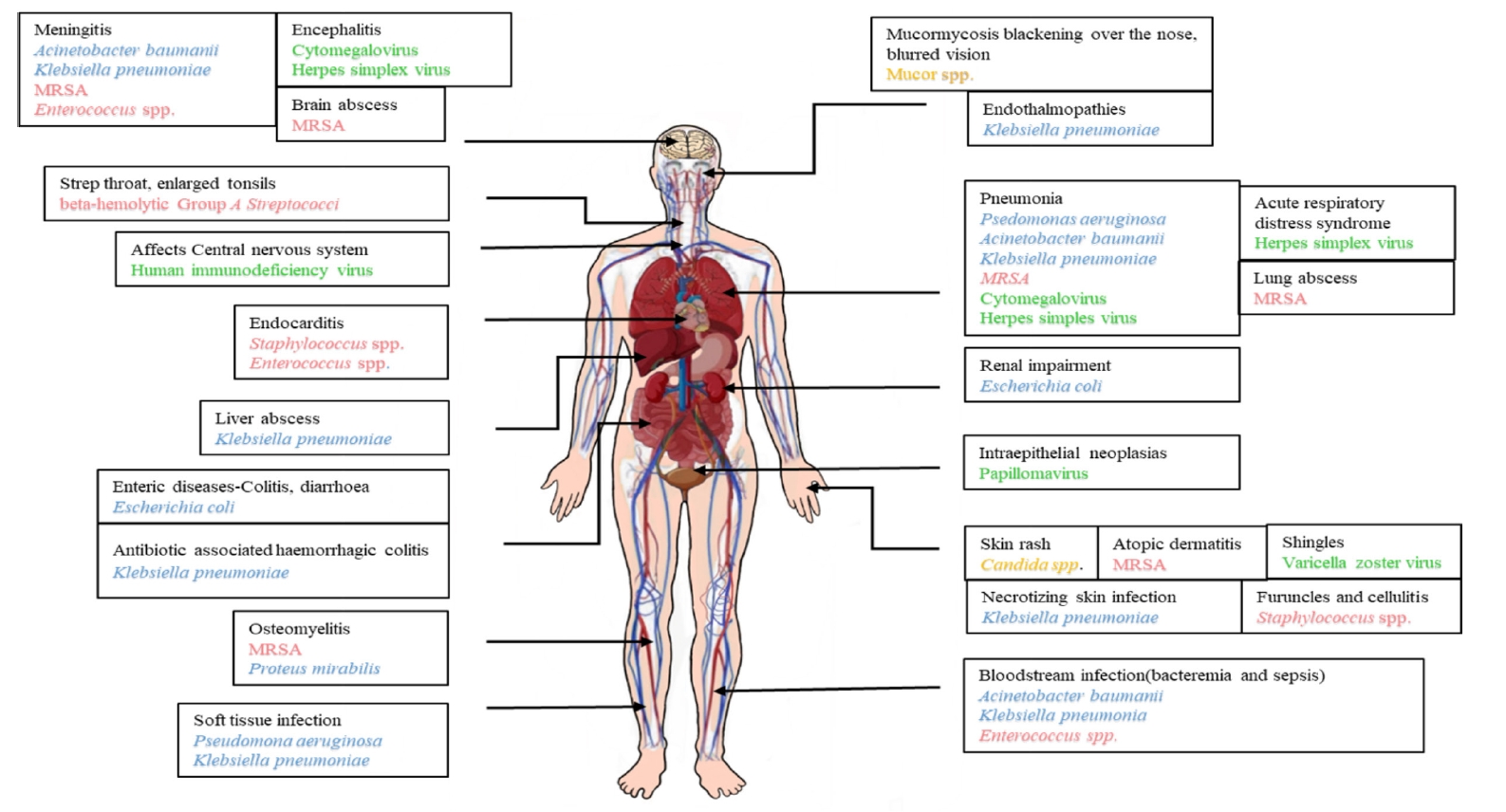
The 2016 National Burn Repository Report mentioned that seven out of ten most frequent complications in burn patients are attributed to polymicrobial burn wound infections (BWIs), with urinary tract infections (UTIs), pneumonia, and cellulitis topping the list and respiratory tract infections being the most frequently reported [13]. After a burn injury, the duration of hospitalization is directly proportional to the types of bacterial species that infect the patients, with the major contributor to infection being Staphylococcus aureus (Figure 1, Table 1) [15]. During the first week of hospitalization, skin and soft tissue infections occur majorly, whereas pneumonia, UTIs, and bloodstream infections tend to occur later during the stay (Table 2) [15].
Gram-Positive Bacteria
The most commonly found Gram-positive bacteria in BWI include Staphylococcus species (spp.), Enterococcus spp., and β-hemolytic group A Streptococci (GAS) [12]. Specifically, vancomycin-resistant Enterococci (VRE) and methicillin-resistant Staphylococcus aureus (MRSA) are the pathogens of high concern in patients with severe burns [12,13]. Over recent decades and with the uncontrolled over-the-counter availability of broad-spectrum antibiotics, MRSA has become the most predominant pathogen in the intensive care unit of burn patients [14]. Colonization with any of these bacteria may also lead to biofilm infections, resulting in severe illness and death [14].
In most of the studies performed so far, about 86.6% of S. aureus found were methicillin-resistant, a major pathogen of hospital-acquired infections (HAIs) in most countries [16]. The toxic products proceeding Staphylococcus spp. infection, such as proteinases, collagenases, and hyaluronidases, allow the bacteria to enter local tissues and the bloodstream, which in turn cause generalized systemic infection and sepsis [14]. In addition to causing pneumonia, sepsis, and other sequelae related to invasive BWIs, Staphylococci are a significant cause of graft loss when the burden of infective organisms exceeds 105 colony-forming units (CFUs) [17]. Vancomycin has been one of the most preferred treatments for curbing MRSA infection. Yet for the past few years, there has been an emergence of other antibiotic-resistant strains like Vancomycin-intermediate Staphylococcus aureus [16]. A potential solution to this problem is being catered to by new antimicrobials such as linezolid (an oxazolidinone), daptomycin, tigecycline, quinupristin-dalfopristin, and dalbavancin [14].
Enterococcus also has been a Gram-positive bacterium of concern but fortunately was not seen to be fatal until the emergence of VRE [18]. Combination therapy, including ampicillin and an aminoglycoside, is nowadays used to treat VRE infections [18]. GAS (Streptococcus pyogenes) is the major cause of graft failure in burn patients, followed by group B Streptococci (Streptococcus agalactiae) [17]. These Streptococci can be eradicated with the penicillin group of antibiotics [19].
Gram-Negative Bacteria
P. aeruginosa are not only the major pathogens that cause respiratory tract infections (HAIs) but are also ubiquitous in invasive burn wounds, owing to their preference for moist environments [20]. These bacteria are also responsible for sepsis, leading to burn-associated death [20]. Pseudomonas infections, particularly those by P. aeruginosa, usually start as a localized, superficial lesion with a typical characteristic yellow or green color and a malodorous fruity smell, which may become an invasive infection termed “ecthyma gangrenosum,” causing blue-purplish “punched-out” lesions in the skin [21]. P. aeruginosa can subsequently spread into deeper tissues rapidly to cause sepsis [22]. Because of the developing drug resistance patterns in P. aeruginosa, piperacillin-tazobactam combination therapy is administered. Aztreonam is used as an alternate therapy for MDR-P. aeruginosa [22].
The Gram-negative bacterium seconding the list of high-concern microbes in burn patients is A. baumannii because of their enhanced capacity for transfer between patients. Survivability in both wet and dry conditions, also on both inanimate and animate objects, helps them to achieve this. [23]. Colistin has been developed as the fallback treatment for pan-resistant Acinetobacter spp. [23].
The failure of burn treatment regimens is mostly caused due to the formation of a biofilm in the burn wound microenvironment of a patient; this may lead to death in many complicated cases [24]. The bacterial community encased within a polysaccharide matrix biofilm is more resistant to disinfection, the rigors of the host immune system, and critically, more tolerant to antibiotics [22]. It is assumed that burn wound-associated biofilms act as a launch pad for the pathogenic bacteria to establish deeper, systemic infections, and ultimately bacteremia and sepsis (Figure 2) [24]. Bacteria of the genera Pseudomonas, Acinetobacter, and Staphylococcus usually adopt a biofilm-encased mode of growth, with P. aeruginosa being the most common (33.3%) burn wound isolate with biofilm-forming abilities, followed by Acinetobacter spp. (23.3%) and Staphylococcus aureus (16.6%) [25,26].
MDR Bacteria
Antibiotics are used as a prophylactic measure to treat burn patients [27]. According to the European Centre for Disease Prevention and Control and the Centers for Disease Control and Prevention, among the drug-resistant (DR) bacteria, there are extensively drug-resistant strains that are resistant to at least one agent in all antimicrobial categories except a few, and pan-drug resistant strains, which are resistant to all agents under all antimicrobial categories [28,29].
Two principal factors that govern MDR-pathogen attacks are the severity and extent of the burn and the duration of hospital stay of the patient [30]. A prolonged hospital stay increases the risk of MDR infections by mostly Gram-negative bacteria (GNB) [30,31]. Further increases in such BWIs might be due to previous exposure to antibiotics, and the use of invasive medical devices like urinary catheters [30]. This was supported by a Canadian Burn Center study, where 125 patients were admitted [32]. Over the first 7 days, 6% of bacterial isolates were MDR, whereas after 28 days of hospital stay, it increased to 44% [32]. This increase in the prevalence of MDR-GNB during long hospital stays of burn patients is thus a serious treatment challenge [33].
Some of the most concerning MDR-GNB strains are A. baumannii, Stenotrophomonas maltophilia, P. aeruginosa, and carbapenem-resistant members of the Enterobacteriaceae family. These, along with Klebsiella pneumoniae, Proteus mirabilis, and Escherichia coli are regarded as the most common MDR-GNB in BWIs [33,34].
In a study conducted at a burn unit of a tertiary care referral center located in North India, it was noted that MRSA and GAS were endemic, where MRSA strains were reported to exhibit resistance to erythromycin, ciprofloxacin, netilmicin, gentamicin, and cefotaxime [35]. MDR P. aeruginosa was also one of the most frequent microbes cultured from the infected burn wounds there, and 90% of those displayed resistance to amikacin and ceftazidime [35].
The preliminary identification of these MDR pathogens is done by studying their physical morphology, Gram-staining properties, and biochemical characteristics [36]. Along with this, antimicrobial susceptibility tests are carried out using various antibiotics, like ciprofloxacin, gentamicin, trimethoprim-sulfamethoxazole, ceftazidime, and others, to check for the zone of growth inhibition [36]. Here, multi-drug resistance is defined if a pathogen shows resistance to at least one agent in 3 or more antimicrobial classes [37].
Yeast and Other Fungal Infections
Fungi are the second major BWI-causing microbes [38]. BWIs caused by fungi are a part of mono- or polymicrobial infections, opportunistic infections, fungemia, and rare aggressive soft tissue infections [39]. These infections are mostly misdiagnosed due to the same kind of manifestations of bacterial infections and due to the lack of a suitable mycology laboratory [38]. These fungal infections have a very high mortality rate, and infection is only nonfatal when there is early diagnosis and treatment [40,41].
From around the globe, 6.3 to 44% of all incident fungal infections have been documented from different burn centers [40,42-44]. From a case study of 220 burn patients, 42% of the BWI pathogens were reported to be Candida spp. [40,42-44]. Invasive Candida infections are one of the major causes of morbidity and mortality among burn patients [42]. Due to the introduction of new antifungals, changes in the epidemiology and drug responses of such fungal infections have been observed [45-48]. It has been found that non-albicans Candida is becoming increasingly resistant to the common anti-mycotic substances [45-49].
Burn patients are usually exposed to these fungal infections after the second week of their thermal injury [50]. The high mortality rate is due to the presence of fungemia, multiple positive cultures, and deep-rooted invasion of healthy skin [51]. The age of the patient, total burn size, body surface area (30%–60%), full-thickness burns, long hospital stay, long-term artificial ventilation, inhalational injury, late surgical excision, artificial dermis, central venous catheters, fungal wound colonization, open dressing, antibiotics (such as imipenem, vancomycin and aminoglycosides), steroid treatment, hyperglycemic episodes, and immunosuppressive disorders all accentuate fungal infections in burn patients [39,45-48,50].
The methods of diagnosis are conventional and mostly organism-specific for the identification of mycoses at the burn site [45]. Direct tissue biopsy is performed in some cases [45]. However due to the voracious growth of fungal culture, sometimes it becomes too late to start an appropriate anti-mycotic therapy [45]. Burn wound samples are collected at proper time intervals for laboratory diagnosis of fungal infections [52]. The burnt tissue should be excised after the 7th, 14th, 21st, and ≥28th days [51]. Tissue biopsy is done for a demonstration of fungal wound infections, and the culture of tissue-specific biopsy is interpreted semi-quantitatively using the following formula [51]:
(1)
CFUs × log reciprocal × 2=colony countTissue weight. (g)
In cultures, the germ tube test, characteristic growth on cornmeal agar, cultural characteristics on HiCrome agar, tetrazolium reduction test, and carbon and nitrogen assimilation tests are evaluated for yeast identification [51]. Molds are identified using lactophenol cotton blue (LPCB) wet mount preparation for conidiogenesis, pattern, and arrangement [51]. Identification of non-sporulating molds is carried out using slide cultures with potato dextrose agar [51].
E-strip or broth micro-dilution using antifungals like amphotericin B, fluconazole, itraconazole, voriconazole, and caspofungin are the tests to check for the antifungal susceptibility of yeasts [52]. The antifungal susceptibility of molds is tested by an E-strip test using amphotericin B [52]. If Candida albicans are isolated, a lower concentration of nystatin is needed as a local treatment in contrast to its higher concentration for the other Candida spp. [40,50,53]. With the burn wounds persisting longer, the propensity of fungal infections increases further [49]. Therefore, the development of pharmaceutical products to recover the wound more rapidly, advancements in topical antifungal therapy, and implementations of appropriate systemic antifungal regimes as guided by antifungal susceptibility tests help to improve the treatment outcomes for severely injured burn patients susceptible to fungal infections [50].
Viral Infections
Burn patients are very susceptible to viral infections [54]. The immunosuppressed state of the patient after an injury triggers the reactivation of latent infection. This becomes the most common cause of viral infection post-injury [54]. Administration of acyclovir for a minimum of 10 days is the most commonly used antiviral therapy to treat viral infection [54].
Herpes simplex virus infections
The frequency of both herpes simplex virus (HSV)-1 and HSV-2 infections in burn patients increases with the age of the victim [54]. There can be primary, secondary, or opportunistic HSV infections due to viral reactivation following reduced immunity in burn patients [54]. It not only impairs the healing process, prolonging the recovery time, but also causes a reduction in the number of T-lymphocytes, down-regulation of Toll-like receptor-mediated nuclear factor-κB expression, and abnormal production of interleukin (IL)-2, cytokines, and antibodies [55,56].
The viral infection manifests itself as groups of vesicopustules or rashes in the burnt area [53]. Reactivation of the latent virus in immune-debilitated burn patients causes diseases like tracheobronchitis, acute respiratory distress syndrome, pneumonia, liver necrosis, focal necrotizing hepatitis, and encephalitis [57].
Fluorescence in-situ hybridization, polymerase chain reaction (PCR), and next generation sequencing are common methods of detecting HSV in BWIs [54]. Intranuclear eosinophilic inclusion bodies in the viral-infected cells are also looked for under a light microscope as a characteristic marker for HSV infections [54].
Cytomegalovirus infections
Burn patients can also be affected by cytomegaloviruses (CMVs), either by primary or exogenous infection or reactivation of latent infections [54]. The infection causes anomalous immune responses involving macrophage hyperactivity, enhanced cytokine production, and over-activation of T-helper cells [58,59]. A 2011 study showed that 71% of CMV infections occurred in CMV-seropositive burn patients, while only 12.5% of CMV-seronegative burn patients were affected [60]. The associated complexities include colitis, pneumonia, organ dysfunction, and encephalitis [60]. PCR, quantitative nucleic acid testing, and immunochemistry are used to detect CMV infections [54]. Histological detection involves the observation of intra-nuclear basophilic inclusion bodies with a characteristic “owl’s-eye” appearance under the light microscope [54].
Varicella zoster virus infections
Varicella zoster virus (VZV) infections in burn injuries are extremely rare, but when they occur, they are accompanied by critical post-infection complications with an increased mortality rate [61]. It is quite prevalent among pediatric burn patients [62]. PCR is the most sensitive method of detecting VZV infections, as compared to culture, serology, or immunochemistry [54]. Sometimes microscopic observation of intranuclear inclusion bodies also confirms the presence of the virus [54]. Any previous infection by the same VZV strains or VZV vaccination lowers the rate of occurrence of VZV infections [62].
Poxvirus infections
Parapoxvirus belonging to the Poxviridae family induces infections in burn patients with skin grafts, either by direct transmission or through infected fomites by indirect transmission [62]. It affects the epidermal keratinocytes of the patients [54]. Vascular endothelial growth factor is upregulated during burn injuries, which promotes angiogenesis, thus facilitating infection [54]. Cell culture isolation, PCR, enzyme-linked immunosorbent assay, and Western blotting are some common methods of detecting the virus [54]. Treatment includes cryotherapy, electrocautery, and the administration of cidofovir or imiquimod [63]. Some large Orf disease (ecthyma contagiosum or contagious pustular dermatitis) lesions might require excision and skin grafting [64].
Human immunodeficiency virus infections
A study of burn patients living with human immunodeficiency virus (HIV) infection in Malawi showed a high probability of death if sepsis or multi-organ dysfunction developed [65]. HIV-positive patients who suffer from burn injury but do not have AIDS are treated similarly to HIV-negative patients [66]. Burn injury, along with a co-existing HIV infection, causes a depletion of CD4+ T cells and defective release of cytokines [67].
Human papillomavirus infections
Human papillomavirus (HPV) replicates when the immune system becomes under-functional in burn patients [54]. These infections were first reported in 1996 when a boy aged 4 years, with a small burn on the left ring finger, was found to develop a “keloid scar” in that burn area, four weeks after the injury [68]. HPV could survive and replicate in the wound, as the basal layer of the skin remained intact [68].
Geographical conditions play a critical role in influencing the development of infection in burn patients, shaping the microbiome found in the BWIs [69,70]. In a study conducted in a hospital in Tanzania, Acinetobacter spp. emerged as the main cause of HAIs in burn patients, whereas in a study done in Nigeria on burn patients, Klebsiella spp. was found to be the predominant pathogen [36,71]. This difference in pathogen preponderance in BWIs is due to varying geographical conditions and different control measures [36]. The survivability of burn patients differs significantly depending on ethnicity and race, as well as on the cost and utilization of health care services [69].
Many countries resorted to social isolation and lockdown for quite a long span of time for the containment of the severe acute respiratory syndrome coronavirus 2 (SARS-CoV-2), which is the causative agent of Coronavirus disease 2019 (COVID-19), the global pandemic. This caused an increase in the occurrence of domestic accidents, leading to burn injuries, although a reduction in amenities available for burn care was observed worldwide during the pandemic, especially in lower-income countries [72].
The skin is the largest anatomical barrier and defensive against the entry of pathogens, which induces a state of immunosuppression when disrupted in burn patients [73]. Host defense has two branches, namely the innate and adaptive immune responses. Of which, the latter takes a longer time to set in [73]. The innate immune response is, however, immediate, severe, and prolonged [73]. At first, there is a pro-inflammatory response where IL-1, IL-6, tumor necrosis factor-α, and interferon-γ cytokines are secreted, and later, the anti-inflammatory response maintains homeostasis by secreting IL-10 and by transforming growth factor-β [73].
Mast cells are the first immune cells to respond to BWIs. Dendritic cells, neutrophils, and monocytes migrate to the site of inflammation under the influence of chemotactic factors [74]. Neutrophils produce reactive oxygen species to destroy the pathogens in the burn wounds, which, in turn, causes damage to skin structures and elicits a strong inflammatory response defined as systemic inflammatory response syndrome (SIRS) [73,75]. SIRS is dampened in elderly patients as compared to younger patients, despite the burn size [73].
The innate immune system is often significantly altered during major burn wounds, where neutrophil and intracellular killings are disrupted, down-regulation of major histocompatibility complex-class II expression occurs, and phagocytic activities of macrophages are diminished [76-78]. These anomalies diminish the natural defenses of the body, increasing the chances of notorious pathogen attacks in burn patients [50,79].
There are three types of BWIs, namely cellulitis, burn wound impetigo, and invasive wound infections within unexcised eschar (necrotizing infection-fasciitis) [80]. Regular laboratory surveillance along with routine microbial wound culturing are essential for strict infection control practices and appropriate antibacterial therapy [80]. Receiving antibiotics before the infection, as well as during the hospitalization period, is a major risk factor for the acquisition of antibiotic-resistant microorganisms [81]. Thus, routine follow-up of the antibiotic-resistance pattern of burn wound flora is absolutely mandatory for successful infection control [81]. Antibiotics must be chosen only after proper monitoring of the antibiotic resistance trend in an individual burn center to restrict infection by MDR microorganisms [80]. Also, systemic antibiotic administration should be carried out for only a very short period of time in burn patients to avoid the spread of multi-drug resistance [81].
Patients with large burn wounds need to be provided with advanced burn wound care [80]. Such advances in wound care include advances in wound exudate and edema control, optimization of the wound environment with ideal skin disinfectants, advances in wound debridement systems, and enhancements in systemic care and management through new applications of medical technologies [82].
Some useful techniques used in burn wound cleansing are high-pressure irrigation, low-pressure irrigation, swabbing, showering, bathing, and washing the affected area under a running liquid [83]. Water, saline, or other antiseptic formulations are used as the cleansing liquid, as applicable [83]. Nowadays, a large number of dressings are available, which are very effective in the healing of cleansed wounds [83]. Some therapeutic applications, involving the use of collagen, hyaluronic acid, growth factor, vacuum-assisted closure, and skin grafting are used to treat burn wounds of varying severities [40]. The Versajet hydrosurgery system is very advantageous for burn wound debridement, which includes optimal preservation of viable tissue, a reduction in blood loss, and effective elimination of bacterial colonization [84].
At the very outset, the prevention of burn injuries should be highly prioritized, as it stands as a global public health crisis, especially in underdeveloped and developing countries. Patients with burn injuries have increased susceptibilities to a wide range of pathogens, including various MDR species of bacteria, fungi, and viruses, particularly during their hospital stay for treatment. This occurs mainly due to their impaired immune system responses, inappropriate vascular organization within the burn-injured area, and intensification of severe oxidative stress. Immunosuppression, prolonged hospitalization, and geographical factors influence the susceptibility of burn patients to MDR-bacterial and fatal viral infections. Microbial transmission and infestation in burn wounds need to be reduced to improve the survival chances of burn patients. For this, an effective infection control policy at every stratum of health care is essential. A combined effort of burn surgeons and burn care units to control the overuse of antibiotics and provide a sterile environment and efficient medical equipment for effective and critical care of the patients should effectively tackle the otherwise sinking situation in burn care across the world.
• The loss of skin epidermis due to burn injury provides easy access for different microorganisms to enter the human body and cause infections.
• Most of the complications related to burn injuries that are reported occur due to the increased susceptibility to several other secondary diseases caused by microbial infections.
• Multidrug-resistant bacterial, yeast, fungal, and viral infections of burn wounds are very common during prolonged hospitalization, and immunosuppression is the main cause.

CONFLICT OF INTEREST

No potential conflict of interest relevant to this article was reported.

FUNDING

None.

ACKNOWLEDGMENTS

The authors express their deepest gratitude to Rev. Dr. Dominic Savio, SJ (Principal of St. Xavier’s College, Kolkata, Autonomous) and Dr. Subhankar Tripathi (Principal of Sarsuna College, Kolkata).

AUTHOR CONTRIBUTIONS

Conceptualization: SR. Data curation: PM, SK, DM, VRC. Methodology: PM, SK, DM, VRC. Writing–original draft: PM, SK, DM, VRC, Writing–review & editing: SR, LC.

Figure 1.
Burn wound infection microbes and their effect on a burn patient. MRSA: methicillin-resistant Staphylococcus aureus; spp.: species.
acc-2023-01571f1.jpg
Figure 2.
(A) Burn wounds typically contain burn wound exudates, which facilitate the initial inoculation and reversible attachment by bacterial pathogens. (B) Bacteria begin to produce extracellular matrix (ECM) and form micro-colonies during the process of irreversible attachment. (C) During the maturation stage, the biofilm grows in size and structural complexity. (D) The mature biofilm enters the dispersal stage, releasing bacterial cells from the ECM, which can then colonize new sites within the wound. Adapted from Maslova et al. NPJ Biofilms Microbiomes 2021;7:73 [3].
acc-2023-01571f2.jpg
Table 1.
Bacterial pathogens isolated from burn wound infections
Bacterial pathogens Percentage of occurrence (%)
Citrobacter freundii 0.77
Escherichia coli 8.46
Klebsiella pneumoniae 13.85
Proteus mirabilis 4.62
Proteus morganii 0.77
Staphylococcus aureus 33.85
Staphylococcus epidermis 3.85
Pseudomonas putida 3.08
Pseudomonas aeruginosa 15.38
Acinetobacter baumannii 15.38

Adapted from El Hamzaoui et al. New Microbes New Infect 2020;38:100764 [15].

Table 2.
Categories and effects of different pathogens causing burn wound infections
Category of microorganisms Microorganisms responsible for causing burn wound infections Prevalence and severity of microorganisms Effect of the microorganism on burn patients
Gram positive bacteria Staphylococcus spp. Most common They cause infection which encompasses causes skin lesions like furuncles, and cellulitis; and sometimes pneumonia, endocarditis, and osteomyelitis, along with biofilm formation.
β-Hemolytic group A Streptococcus Common They cause strep throat, enlarged lymph nodes in the neck, enlarged tonsils and rash.
Enterococcus spp. Common They cause bacteremia, and infective endocarditis, UTIs, meningitis, and rarely causes intra-abdominal infections.
Gram negative bacteria Acinetobacter baumannii Most common, dangerous It causes diseases such as pneumonia and meningitis, bloodstream infections (bacteremia and sepsis), delays in wound healing, graft losses, UTIs.
Klebsiella pneumoniae Most common It causes endophthalmitis, pyrogenic liver abscess, splenic abscess, necrotizing skin infection, soft tissue infection, meningitis, antibiotic-associated hemorrhagic colitis, bacteremia, pneumonia, Lemierre syndrome.
Pseudomonas aeruginosa Most common, concerning It causes infections in the blood, lungs (pneumonia), soft tissue infection, UTIs.
Escherichia coli Common It causes enteric diseases, such as diarrhoea/dysentery, colitis, meningitis, low grade fever, vomiting, renal impairment.
Multidrug resistant bacteria P. aeruginosa Most common, dangerous It causes infections in the blood, lungs (pneumonia), soft tissue infection, UTIs.
A. baumannii Most common It causes diseases such as pneumonia and meningitis, bloodstream infections (bacteremia and sepsis), delays in wound healing, graft losses, UTIs.
Klebsiella pneumoniae Most common, concerning It causes endophthalmitis, pyrogenic liver abscess, splenic abscess, necrotizing skin infection, soft tissue infection, meningitis, antibiotic-associated hemorrhagic colitis, bacteremia, pneumonia, Lemierre syndrome.
Methicillin-resistant Staphylococcus aureus Common, dangerous MRSA causes skin infections like atopic dermatitis, followed by invasive infections like osteomyelitis, meningitis, lung abscess, pneumonia, brain abscess and central nervous system infection.
Escherichia coli Common It causes enteric diseases, such as diarrhoea/dysentery, colitis, meningitis, low grade fever, vomiting, renal impairment.
Proteus mirabilis Common It mostly causes UTIs, along with meningoencephalitis, empyema, and osteomyelitis.
Fungi Candida spp. Most common They cause intense itching. Symptoms also include red, growing skin rash, rash on the skin folds, genitals, middle of the body, buttocks, under the breasts, and other areas of skin.
Aspergillus fumigatus Most common It causes infections usually in people who have weakened immune systems.
Saccharomyces boulardii Uncommon It causes fungemia.
Mucor spp. Uncommon, dangerous They cause mucormycosis; fatal.
Viruses Herpes simplex virus Most common, very dangerous It affects production of antibodies, cytokines, T-cells, IL-2, etc. Reactivation of the virus causes acute respiratory distress syndrome, pneumonia, liver necrosis, and encephalitis.
Cytomegalovirus Most common, very dangerous It increases production of cytokines and causes hyperactivity of T helper cells and macrophages. It leads to organ dysfunction, pneumonia, encephalitis, and colitis.
Varicella zoster virus Common, dangerous It causes shingles; post-herpetic neuralgia and delayed healing.
Poxvirus Rare It causes formation of lesions and scabs.
Human immunodeficiency virus Rare It decreases population of CD4+ T-cells. It eventually leads to chronic multi-organ diseases and severe impairments within the central nervous system.
Papillomavirus Rare It causes intraepithelial neoplasias.

spp.: species; UTI: urinary tract infection; MRSA: Methicillin-resistant Staphylococcus aureus; IL: interleukin.

  • 1. Mohapatra S, Gupta A, Agrawal K. Bacteriological profiles in burn patients within first twenty-four hours of injury. Int J Med Microbiol Trop Dis 2016;2:71-4.Article
  • 2. Chen YY, Wu PF, Chen CS, Chen IH, Huang WT, Wang FD. Trends in microbial profile of burn patients following an event of dust explosion at a tertiary medical center. BMC Infect Dis 2020;20:193. ArticlePubMedPMCPDF
  • 3. Maslova E, Eisaiankhongi L, Sjöberg F, McCarthy RR. Burns and biofilms: priority pathogens and in vivo models. NPJ Biofilms Microbiomes 2021;7:73. ArticlePubMedPMCPDF
  • 4. López-Jácome LE, Chávez-Heres T, Becerra-Lobato N, García-Hernández ML, Vanegas-Rodríguez ES, Colin-Castro CA, et al. Microbiology and infection profile of electric burned patients in a referral burn hospital in Mexico City. J Burn Care Res 2020;41:390-7.ArticlePubMedPDF
  • 5. Forjuoh SN. Burns in low- and middle-income countries: a review of available literature on descriptive epidemiology, risk factors, treatment, and prevention. Burns 2006;32:529-37.ArticlePubMed
  • 6. Peck MD. Epidemiology of burns throughout the world. Part I: distribution and risk factors. Burns 2011;37:1087-100.ArticlePubMed
  • 7. Ho WS, Ying SY. An epidemiological study of 1063 hospitalized burn patients in a tertiary burns centre in Hong Kong. Burns 2001;27:119-23.ArticlePubMed
  • 8. Opriessnig E, Luze H, Smolle C, Draschl A, Zrim R, Giretzlehner M, et al. Epidemiology of burn injury and the ideal dressing in global burn care: regional differences explored. Burns 2023;49:1-14.ArticlePubMed
  • 9. Peck MD. Epidemiology of burns throughout the World. Part II: intentional burns in adults. Burns 2012;38:630-7.ArticlePubMed
  • 10. Kara YA. Hot topics in burn injuries. Chapter 2: burn etiology and pathogenesis. Intechope. 2018.
  • 11. Elsous A, Ouda M, Mohsen S, Al-Shaikh M, Mokayad S, Abo-Shaban N, et al. Epidemiology and outcomes of hospitalized burn patients in Gaza strip: a descriptive study. Ethiop J Health Sci 2016;26:9-16.ArticlePubMedPMC
  • 12. Bhardwaj S, Bhatia S, Singh S, Franco F Jr. Growing emergence of drug-resistant Pseudomonas aeruginosa and attenuation of its virulence using quorum sensing inhibitors: a critical review. Iran J Basic Med Sci 2021;24:699-719.PubMedPMC
  • 13. Jeschke MG, van Baar ME, Choudhry MA, Chung KK, Gibran NS, Logsetty S. Burn injury. Nat Rev Dis Primers 2020;6:11. ArticlePubMedPMCPDF
  • 14. Norbury W, Herndon DN, Tanksley J, Jeschke MG, Finnerty CC. Infection in burns. Surg Infect (Larchmt) 2016;17:250-5.ArticlePubMedPMC
  • 15. El Hamzaoui N, Barguigua A, Larouz S, Maouloua M. Epidemiology of burn wound bacterial infections at a Meknes hospital, Morocco. New Microbes New Infect 2020;38:100764. ArticlePubMedPMC
  • 16. Thabet L, Messadi Aa, Mbarek M, Turki A, Meddeb B, Ben Redjeb S. Surveillance of multidrug resistant bacteria in a Tunisian hospital. Tunis Med 2008;86:992-5.PubMed
  • 17. Thabet L, Turki A, Ben Redjeb S, Messadi A. Bacteriological profile and antibiotic resistance of bacteria isolates in a burn department. Tunis Med 2008;86:1051-4.ArticlePubMed
  • 18. von Baum H, Ober JF, Wendt C, Wenzel RP, Edmond MB. Antibiotic-resistant bloodstream infections in hospitalized patients: specific risk factors in a high-risk population? Infection 2005;33:320-6.ArticlePubMedPDF
  • 19. Wilson GR, French GW, Sully L. Loss of split thickness skin grafts due to non-group A beta-haemolytic streptococci. Ann R Coll Surg Engl 1988;70:217-9.PubMedPMC
  • 20. Williams FN, Herndon DN, Hawkins HK, Lee JO, Cox RA, Kulp GA, et al. The leading causes of death after burn injury in a single pediatric burn center. Crit Care 2009;13:R183. ArticlePubMedPMC
  • 21. McManus AT, Mason AD, McManus WF, Pruitt BA. Twenty-five year review of Pseudomonas aeruginosa bacteremia in a burn center. Eur J Clin Microbiol 1985;4:219-23.ArticlePubMedPDF
  • 22. Walton MA, Villarreal C, Herndon DN, Heggers JP. The use of aztreonam as an alternate therapy for multi-resistant Pseudomonas aeruginosa. Burns 1997;23:225-7.ArticlePubMed
  • 23. Corbella X, Montero A, Pujol M, Domínguez MA, Ayats J, Argerich MJ, et al. Emergence and rapid spread of carbapenem resistance during a large and sustained hospital outbreak of multiresistant Acinetobacter baumannii. J Clin Microbiol 2000;38:4086-95.ArticlePubMedPMCPDF
  • 24. Thomas RE, Thomas BC. Reducing biofilm infections in burn patients' wounds and biofilms on surfaces in hospitals, medical facilities and medical equipment to improve burn care: a systematic review. Int J Environ Res Public Health 2021;18:13195. ArticlePubMedPMC
  • 25. Khan BA, Yeh AJ, Cheung GY, Otto M. Investigational therapies targeting quorum-sensing for the treatment of Staphylococcus aureus infections. Expert Opin Investig Drugs 2015;24:689-704.ArticlePubMedPMC
  • 26. Ramakrishnan M, Putli Bai S, Babu M. Study on biofilm formation in burn wound infection in a pediatric hospital in Chennai, India. Ann Burns Fire Disasters 2016;29:276-80.PubMedPMC
  • 27. Avni T, Levcovich A, Ad-El DD, Leibovici L, Paul M. Prophylactic antibiotics for burns patients: systematic review and meta-analysis. BMJ 2010;340:c241. ArticlePubMedPMC
  • 28. Falagas ME, Koletsi PK, Bliziotis IA. The diversity of definitions of multidrug-resistant (MDR) and pandrug-resistant (PDR) Acinetobacter baumannii and Pseudomonas aeruginosa. J Med Microbiol 2006;55(Pt 12):1619-29.ArticlePubMed
  • 29. Gupta M, Naik AK, Singh SK. Bacteriological profile and antimicrobial resistance patterns of burn wound infections in a tertiary care hospital. Heliyon 2019;5:e02956. ArticlePubMedPMC
  • 30. BioMérieux. Burn patients often have higher rates of multidrug-resistant infections—but there are ways to help [Internet]. BioMérieux. 2023;[cited 2024 May 1]. Available from: https://www.biomerieux.com/nl/en/blog/antimicrobial-resistance-stewardship/Burn-Patients-Multidrug-Resistant-Infections.html.
  • 31. Lachiewicz AM, Hauck CG, Weber DJ, Cairns BA, van Duin D. Bacterial infections after burn injuries: impact of multidrug resistance. Clin Infect Dis 2017;65:2130-6.ArticlePubMedPMC
  • 32. Wanis M, Walker SA, Daneman N, Elligsen M, Palmay L, Simor A, et al. Impact of hospital length of stay on the distribution of Gram negative bacteria and likelihood of isolating a resistant organism in a Canadian burn center. Burns 2016;42:104-11.ArticlePubMed
  • 33. Sheridan R, Weber J, Chang P. Multi-drug resistant Gram-negative bacteria colonization and infection in burned children: lessons learned from a 20-year experience. Burns Open 2018;2:43-6.Article
  • 34. Rosanova MT, Stamboulian D, Lede R. Risk factors for mortality in burn children. Braz J Infect Dis 2014;18:144-9.ArticlePubMed
  • 35. Taneja N, Emmanuel R, Chari PS, Sharma M. A prospective study of hospital-acquired infections in burn patients at a tertiary care referral centre in North India. Burns 2004;30:665-9.ArticlePubMed
  • 36. Kabanangi F, Joachim A, Nkuwi EJ, Manyahi J, Moyo S, Majigo M. High level of multidrug-resistant gram-negative pathogens causing burn wound infections in hospitalized children in Dar es Salaam, Tanzania. Int J Microbiol 2021;2021:6644185. ArticlePubMedPMCPDF
  • 37. Magiorakos AP, Srinivasan A, Carey RB, Carmeli Y, Falagas ME, Giske CG, et al. Multidrug-resistant, extensively drug-resistant and pandrug-resistant bacteria: an international expert proposal for interim standard definitions for acquired resistance. Clin Microbiol Infect 2012;18:268-81.ArticlePubMed
  • 38. Gupta N, Haque A, Lattif AA, Narayan RP, Mukhopadhyay G, Prasad R. Epidemiology and molecular typing of Candida isolates from burn patients. Mycopathologia 2004;158:397-405.ArticlePubMedPDF
  • 39. Horvath EE, Murray CK, Vaughan GM, Chung KK, Hospenthal DR, Wade CE, et al. Fungal wound infection (not colonization) is independently associated with mortality in burn patients. Ann Surg 2007;245:978-85.ArticlePubMedPMC
  • 40. Mousa HA. Fungal infection of burn wounds in patients with open and occlusive treatment methods. East Mediterr Health J 1999;5:333-6.ArticlePubMed
  • 41. Ahmad S, Khan Z, Mustafa AS, Khan ZU. Epidemiology of Candida colonization in an intensive care unit of a teaching hospital in Kuwait. Med Mycol 2003;41:487-93.ArticlePubMed
  • 42. Murray CK, Loo FL, Hospenthal DR, Cancio LC, Jones JA, Kim SH, et al. Incidence of systemic fungal infection and related mortality following severe burns. Burns 2008;34:1108-12.ArticlePubMed
  • 43. Becker WK, Cioffi WG, McManus AT, Kim SH, McManus WF, Mason AD, et al. Fungal burn wound infection: a 10-year experience. Arch Surg 1991;126:44-8.ArticlePubMed
  • 44. Ballard J, Edelman L, Saffle J, Sheridan R, Kagan R, Bracco D, et al. Positive fungal cultures in burn patients: a multicenter review. J Burn Care Res 2008;29:213-21.ArticlePubMed
  • 45. Greenhalgh DG, Saffle JR, Holmes JH, Gamelli RL, Palmieri TL, Horton JW, et al. American Burn Association consensus conference to define sepsis and infection in burns. J Burn Care Res 2007;28:776-90.ArticlePubMed
  • 46. Mathew BP, Nath M. Recent approaches to antifungal therapy for invasive mycoses. ChemMedChem 2009;4:310-23.ArticlePubMed
  • 47. Dries DJ. Management of burn injuries: recent developments in resuscitation, infection control and outcomes research. Scand J Trauma Resusc Emerg Med 2009;17:14. ArticlePubMedPMC
  • 48. Struck MF. Infection control in burn patients: are fungal infections underestimated? Scand J Trauma Resusc Emerg Med 2009;17:51-6.ArticlePubMedPMC
  • 49. de Macedo JL, Santos JB. Bacterial and fungal colonization of burn wounds. Mem Inst Oswaldo Cruz 2005;100:535-9.ArticlePubMed
  • 50. Church D, Elsayed S, Reid O, Winston B, Lindsay R. Burn wound infections. Clin Microbiol Rev 2006;19:403-34.ArticlePubMedPMCPDF
  • 51. Capoor MR, Sarabahi S, Tiwari VK, Narayanan RP. Fungal infections in burns: diagnosis and management. Indian J Plast Surg 2010;43(Suppl):S37-42.ArticlePubMedPMC
  • 52. National Committee for Clinical Laboratory Standards. Reference method for broth dilution antifungal susceptibility testing of yeasts; approved standard. Second edition. NCCLS document M27-A2 [ISBN 1-56238-469-4] [Internet]. National Committee for Clinical Laboratory Standards. 2002;[cited 2024 May 1]. Available from: https://webstore.ansi.org/preview-pages/CLSI/preview_M27-A2.pdf.
  • 53. Mousa HA. Aerobic, anaerobic and fungal burn wound infections. J Hosp Infect 1997;37:317-23.ArticlePubMed
  • 54. Baj J, Korona-Głowniak I, Buszewicz G, Forma A, Sitarz M, Teresiński G. Viral infections in burn patients: a state-of-the-art review. Viruses 2020;12:1315. ArticlePubMedPMC
  • 55. Xu H, Su C, Pearson A, Mody CH, Zheng C. Herpes simplex virus 1 UL24 abrogates the DNA sensing signal pathway by inhibiting NF-κB activation. J Virol 2017;91:e00025-17.ArticlePubMedPMCPDF
  • 56. Wood JJ, O'Mahony JB, Rodrick ML, Eaton R, Demling RH, Mannick JA. Abnormalities of antibody production after thermal injury: an association with reduced interleukin 2 production. Arch Surg 1986;121:108-15.ArticlePubMed
  • 57. Haik J, Weissman O, Stavrou D, Ben-noon HI, Liran A, Tessone A, et al. Is prophylactic acyclovir treatment warranted for prevention of herpes simplex virus infections in facial burns? A review of the literature. J Burn Care Res 2011;32:358-62.ArticlePubMed
  • 58. Rennekampff HO, Hamprecht K. Cytomegalovirus infection in burns: a review. J Med Microbiol 2006;55:483-7.ArticlePubMed
  • 59. Schwacha MG. Macrophages and post-burn immune dysfunction. Burns 2003;29:1-14.ArticlePubMed
  • 60. Bordes J, Maslin J, Prunet B, d'Aranda E, Lacroix G, Goutorbe P, et al. Cytomegalovirus infection in severe burn patients monitoring by real-time polymerase chain reaction: a prospective study. Burns 2011;37:434-9.ArticlePubMed
  • 61. Wurzer P, Guillory A, Parvizi D, Clayton RP, Branski LK, Kamolz LP, et al. Human herpes viruses in burn patients: a systematic review. Burns 2017;43:25-33.ArticlePubMed
  • 62. Sheridan RL, Weber JM, Pasternak MM, Mulligan JM, Tompkins RG. A 15-year experience with varicella infections in a pediatric burn unit. Burns 1999;25:353-6.ArticlePubMed
  • 63. Midilli K, Erkiliç A, Kuşkucu M, Analay H, Erkiliç S, Benzonana N, et al. Nosocomial outbreak of disseminated orf infection in a burn unit, Gaziantep, Turkey, October to December 2012. Euro Surveill 2013;18:20425. ArticlePubMed
  • 64. Bergqvist C, Kurban M, Abbas O. Orf virus infection. Rev Med Virol 2017;27.ArticlePDF
  • 65. Al-Qattan MM. Orf infection of the hand. J Hand Surg Am 2011;36:1855-8.ArticlePubMed
  • 66. Edge JM, Van der Merwe AE, Pieper CH, Bouic P. Clinical outcome of HIV positive patients with moderate to severe burns. Burns 2001;27:111-4.ArticlePubMed
  • 67. Allgöwer M, Schoenenberger GA, Sparkes BG. Burning the largest immune organ. Burns 1995;21 Suppl 1:S7-47.PubMed
  • 68. Camilleri IG, Milner RH. Human papilloma virus proliferation in a healing burn. Burns 1996;22:162-3.ArticlePubMed
  • 69. Espinoza LF, Friedstat J, Faoro N, Chang PH, McMullen KA, Simko LC, et al. Geographic variation in outcomes after burn injury: a burn model system national database study. Ann Plast Surg 2020;84:644-50.ArticlePubMedPMC
  • 70. Gupta VK, Paul S, Dutta C. Geography, ethnicity or subsistence-specific variations in human microbiome composition and diversity. Front Microbiol 2017;8:1162. ArticlePubMedPMC
  • 71. Ozumba UC, Jiburum BC. Bacteriology of burn wounds in Enugu, Nigeria. Burns 2000;26:178-80.ArticlePubMed
  • 72. Laura P, José A, Nikki A, Khaled A, Barret J, Jeffery C, et al. Impact of COVID-19 on global burn care. Burns 2022;48:1301-10.ArticlePubMed
  • 73. Sierawska O, Małkowska P, Taskin C, Hrynkiewicz R, Mertowska P, Grywalska E, et al. Innate immune system response to burn damage-focus on cytokine alteration. Int J Mol Sci 2022;23:716. ArticlePubMedPMC
  • 74. Vinish M, Cui W, Stafford E, Bae L, Hawkins H, Cox R, et al. Dendritic cells modulate burn wound healing by enhancing early proliferation. Wound Repair Regen 2016;24:6-13.ArticlePubMed
  • 75. Rani M, Schwacha MG. Aging and the pathogenic response to burn. Aging Dis 2012;3:171-80.PubMed
  • 76. Grogan JB. Altered neutrophil phagocytic function in burn patients. J Trauma 1976;16:734-8.ArticlePubMed
  • 77. Rich RR. Clinical immunology principles and practices. 2001, Mosby.
  • 78. Altman LC, Furukawa CT, Klebanoff SJ. Depressed mononuclear leukocyte chemotaxis in thermally injured patients. J Immunol 1977;119:199-205.ArticlePubMedPDF
  • 79. Moins-Teisserenc H, Cordeiro DJ, Audigier V, Ressaire Q, Benyamina M, Lambert J, et al. Severe altered immune status after burn injury is associated with bacterial infection and septic shock. Front Immunol 2021;12:586195. ArticlePubMedPMC
  • 80. Coban YK. Infection control in severely burned patients. World J Crit Care Med 2012;1:94-101.ArticlePubMedPMC
  • 81. Boyce JM, White RL, Causey WA, Lockwood WR. Burn units as a source of methicillin-resistant Staphylococcus aureus infections. JAMA 1983;249:2803-7.ArticlePubMed
  • 82. Wolvos T. Wound instillation: the next step in negative pressure wound therapy. Lessons learned from initial experiences. Ostomy Wound Manage 2004;50:56-66.
  • 83. Wilson JR, Mills JG, Prather ID, Dimitrijevich SD. A toxicity index of skin and wound cleansers used on in vitro fibroblasts and keratinocytes. Adv Skin Wound Care 2005;18:373-8.ArticlePubMed
  • 84. Coban YK, Erkiliç A, Analay H. Our 18-month experience at a new burn center in Gaziantep, Turkey. Ulus Travma Acil Cerrahi Derg 2010;16:353-6.PubMed

Figure & Data

References

    Citations

    Citations to this article as recorded by  

      • PubReader PubReader
      • ePub LinkePub Link
      • Cite
        CITE
        export Copy
        Close
        Download Citation
        Download a citation file in RIS format that can be imported by all major citation management software, including EndNote, ProCite, RefWorks, and Reference Manager.

        Format:
        • RIS — For EndNote, ProCite, RefWorks, and most other reference management software
        • BibTeX — For JabRef, BibDesk, and other BibTeX-specific software
        Include:
        • Citation for the content below
        Microbial infections in burn patients
        Acute Crit Care. 2024;39(2):214-225.   Published online May 24, 2024
        Close
      • XML DownloadXML Download
      Figure
      We recommend

      ACC : Acute and Critical Care